 Movie
Movie Controller

[English] 日本語
 Yorodumi
- PDB-8jnp: Crystal structure of cytochrome P450 CftA from Streptomyces torul... -
+ Open data

- Basic information
| Entry | Database: PDB / ID: 8jnp | |||||||||
|---|---|---|---|---|---|---|---|---|---|---|
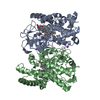
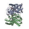
| Title | Crystal structure of cytochrome P450 CftA from Streptomyces torulosus NRRL B-3889, in complex with the substrate ikarugamycin | |||||||||
 Components Components | Cytochrome P450 CftA | |||||||||
 Keywords Keywords | OXIDOREDUCTASE / Actinomadura viridis / hydrolase / Lobophorin / deglycosylation | |||||||||
| Function / homology | Chem-EIA / PROTOPORPHYRIN IX CONTAINING FE Function and homology information Function and homology information | |||||||||
| Biological species |  Streptomyces torulosus (bacteria) Streptomyces torulosus (bacteria) | |||||||||
| Method |  X-RAY DIFFRACTION / MOLECULAR REPLACEMENT / Resolution: 2 Å X-RAY DIFFRACTION / MOLECULAR REPLACEMENT / Resolution: 2 Å | |||||||||
 Authors Authors | Jiang, P. / Zhang, L.P. / Zhang, C.S. | |||||||||
| Funding support |  China, 2items China, 2items
| |||||||||
 Citation Citation | Journal: Angew.Chem.Int.Ed.Engl. / Year: 2023 Title: A Mechanistic Understanding of the Distinct Regio- and Chemoselectivity of Multifunctional P450s by Structural Comparison of IkaD and CftA Complexed with Common Substrates. Authors: Jiang, P. / Jin, H. / Zhang, G. / Zhang, W. / Liu, W. / Zhu, Y. / Zhang, C. / Zhang, L. | |||||||||
| History |
|
- Structure visualization
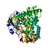
| Structure viewer | Molecule: MolmilJmol/JSmol |
|---|
- Downloads & links
-Download
| PDBx/mmCIF format | 8jnp.cif.gz | 171.2 KB | Display | PDBx/mmCIF format |
|---|---|---|---|---|
| PDB format | pdb8jnp.ent.gz | 134 KB | Display | PDB format |
| PDBx/mmJSON format | 8jnp.json.gz | Tree view | PDBx/mmJSON format | |
| Others |  Other downloads Other downloads |
-Validation report
| Arichive directory | https://data.pdbj.org/pub/pdb/validation_reports/jn/8jnpftp://data.pdbj.org/pub/pdb/validation_reports/jn/8jnp | HTTPS FTP |
|---|
-Related structure data

-Links
 PDBj
PDBj- Assembly
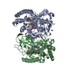
| Deposited unit | 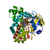
| ||||||||
|---|---|---|---|---|---|---|---|---|---|
| 1 |
| ||||||||
| Unit cell |
|
-Components
| #1: Protein | Mass: 44088.637 Da / Num. of mol.: 1 Source method: isolated from a genetically manipulated source Source: (gene. exp.) Streptomyces torulosus (bacteria) / Strain: NRRL B-3889 / Production host: |
|---|---|
| #2: Chemical | ChemComp-EIA / (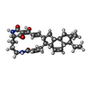  Mass: 478.623 Da / Num. of mol.: 1 / Source method: obtained synthetically / Formula: C29H38N2O4 / Feature type: SUBJECT OF INVESTIGATION Mass: 478.623 Da / Num. of mol.: 1 / Source method: obtained synthetically / Formula: C29H38N2O4 / Feature type: SUBJECT OF INVESTIGATION |
| #3: Chemical | ChemComp-HEM /   Mass: 616.487 Da / Num. of mol.: 1 / Source method: obtained synthetically / Formula: C34H32FeN4O4 / Feature type: SUBJECT OF INVESTIGATION Mass: 616.487 Da / Num. of mol.: 1 / Source method: obtained synthetically / Formula: C34H32FeN4O4 / Feature type: SUBJECT OF INVESTIGATION |
| #4: Water | ChemComp-HOH /  Mass: 18.015 Da / Num. of mol.: 388 / Source method: isolated from a natural source / Formula: H2O Mass: 18.015 Da / Num. of mol.: 388 / Source method: isolated from a natural source / Formula: H2O |
| Has ligand of interest | Y |
-Experimental details
-Experiment
| Experiment | Method: X-RAY DIFFRACTION / Number of used crystals: 1 |
|---|
- Sample preparation
| Crystal | Density Matthews: 2.83 Å3/Da / Density % sol: 56.61 % |
|---|---|
| Crystal grow | Temperature: 289 K / Method: vapor diffusion, sitting drop / pH: 6 / Details: 0.2M sodium chloride, 0.1M MES, 20% W/V PEG 6000 |
-Data collection
| Diffraction | Mean temperature: 100 K / Serial crystal experiment: N |
|---|---|
| Diffraction source | Source: ROTATING ANODE / Type: RIGAKU / Wavelength: 1.54184 Å |
| Detector | Type: DECTRIS PILATUS 200K / Detector: PIXEL / Date: Sep 22, 2021 |
| Radiation | Protocol: SINGLE WAVELENGTH / Monochromatic (M) / Laue (L): M / Scattering type: x-ray |
| Radiation wavelength | Wavelength: 1.54184 Å / Relative weight: 1 |
| Reflection | Resolution: 2→19.06 Å / Num. obs: 34584 / % possible obs: 99.9 % / Redundancy: 10.5 % / CC1/2: 0.998 / Rmerge(I) obs: 0.106 / Rpim(I) all: 0.033 / Rrim(I) all: 0.111 / Χ2: 0.97 / Net I/σ(I): 13.5 / Num. measured all: 362272 |
| Reflection shell | Resolution: 2→2.05 Å / % possible obs: 100 % / Redundancy: 7.2 % / Rmerge(I) obs: 0.559 / Num. measured all: 18120 / Num. unique obs: 2504 / CC1/2: 0.926 / Rpim(I) all: 0.222 / Rrim(I) all: 0.602 / Χ2: 0.48 / Net I/σ(I) obs: 2.3 |
- Processing
| Software |
| |||||||||||||||||||||||||||||||||||||||||||||||||||||||||||||||||||||||||||||||||||||||||||
|---|---|---|---|---|---|---|---|---|---|---|---|---|---|---|---|---|---|---|---|---|---|---|---|---|---|---|---|---|---|---|---|---|---|---|---|---|---|---|---|---|---|---|---|---|---|---|---|---|---|---|---|---|---|---|---|---|---|---|---|---|---|---|---|---|---|---|---|---|---|---|---|---|---|---|---|---|---|---|---|---|---|---|---|---|---|---|---|---|---|---|---|---|
| Refinement | Method to determine structure: MOLECULAR REPLACEMENT / Resolution: 2→19.06 Å / SU ML: 0.22 / Cross valid method: FREE R-VALUE / σ(F): 1.33 / Phase error: 24.57 / Stereochemistry target values: ML
| |||||||||||||||||||||||||||||||||||||||||||||||||||||||||||||||||||||||||||||||||||||||||||
| Solvent computation | Shrinkage radii: 0.9 Å / VDW probe radii: 1.11 Å / Solvent model: FLAT BULK SOLVENT MODEL | |||||||||||||||||||||||||||||||||||||||||||||||||||||||||||||||||||||||||||||||||||||||||||
| Refinement step | Cycle: LAST / Resolution: 2→19.06 Å
| |||||||||||||||||||||||||||||||||||||||||||||||||||||||||||||||||||||||||||||||||||||||||||
| Refine LS restraints |
| |||||||||||||||||||||||||||||||||||||||||||||||||||||||||||||||||||||||||||||||||||||||||||
| LS refinement shell |
|
