 Movie
Movie Controller

[English] 日本語
 Yorodumi
- PDB-8jfn: Crystal structure of enoyl-ACP reductase FabI in complex with NAD... -
+ Open data

- Basic information
| Entry | Database: PDB / ID: 8jfn | ||||||
|---|---|---|---|---|---|---|---|
| Title | Crystal structure of enoyl-ACP reductase FabI in complex with NAD+ and crotonyl-ACP from Helicobacter pylori | ||||||
 Components Components |
| ||||||
 Keywords Keywords | BIOSYNTHETIC PROTEIN / enoyl-ACP reductase / FabI / NAD+ / crotonyl-ACP / Helicobacter pylori | ||||||
| Function / homology |  Function and homology information Function and homology informationKdo2-lipid A biosynthetic process / enoyl-[acyl-carrier-protein] reductase (NADH) / enoyl-[acyl-carrier-protein] reductase (NADH) activity / lipid A biosynthetic process / phosphopantetheine binding / acyl binding / acyl carrier activity / fatty acid biosynthetic process / nucleotide binding / membrane / cytosol Similarity search - Function | ||||||
| Biological species |   Helicobacter pylori (bacteria) Helicobacter pylori (bacteria) | ||||||
| Method |  X-RAY DIFFRACTION / SYNCHROTRON / MOLECULAR REPLACEMENT / Resolution: 2.41 Å X-RAY DIFFRACTION / SYNCHROTRON / MOLECULAR REPLACEMENT / Resolution: 2.41 Å | ||||||
 Authors Authors | Song, W.Y. / Zhang, L. | ||||||
| Funding support |  China, 1items China, 1items
| ||||||
 Citation Citation | Journal: Angew.Chem.Int.Ed.Engl. / Year: 2023 Title: The Molecular Basis of Catalysis by SDR Family Members Ketoacyl-ACP Reductase FabG and Enoyl-ACP Reductase FabI in Type-II Fatty Acid Biosynthesis. Authors: Zhou, J. / Zhang, L. / Wang, Y. / Song, W. / Huang, Y. / Mu, Y. / Schmitz, W. / Zhang, S.Y. / Lin, H. / Chen, H.Z. / Ye, F. / Zhang, L. | ||||||
| History |
|
- Structure visualization
| Structure viewer | Molecule: MolmilJmol/JSmol |
|---|
- Downloads & links
-Download
| PDBx/mmCIF format | 8jfn.cif.gz | 86.1 KB | Display | PDBx/mmCIF format |
|---|---|---|---|---|
| PDB format | pdb8jfn.ent.gz | 62.4 KB | Display | PDB format |
| PDBx/mmJSON format | 8jfn.json.gz | Tree view | PDBx/mmJSON format | |
| Others |  Other downloads Other downloads |
-Validation report
| Arichive directory | https://data.pdbj.org/pub/pdb/validation_reports/jf/8jfnftp://data.pdbj.org/pub/pdb/validation_reports/jf/8jfn | HTTPS FTP |
|---|
-Related structure data
| Related structure data |  8jf9C  8jfaC  8jfgC  8jfhC  8jfiC  8jfjC  8jfmSC S: Starting model for refinement C: citing same article ( |
|---|---|
| Similar structure data |
-Links
 PDBj
PDBj

- Assembly
| Deposited unit | 
| ||||||||||||
|---|---|---|---|---|---|---|---|---|---|---|---|---|---|
| 1 | 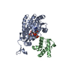
| ||||||||||||
| Unit cell |
| ||||||||||||
| Components on special symmetry positions |
|
-Components
| #1: Protein | Mass: 29746.154 Da / Num. of mol.: 1 Source method: isolated from a genetically manipulated source Source: (gene. exp.) Helicobacter pylori (bacteria) / Gene: fabI / Production host: |
|---|---|
| #2: Protein | Mass: 8740.794 Da / Num. of mol.: 1 Source method: isolated from a genetically manipulated source Source: (gene. exp.) Helicobacter pylori (bacteria) / Gene: acpP / Production host: |
| #3: Chemical | ChemComp-NAD /   Mass: 663.425 Da / Num. of mol.: 1 / Source method: obtained synthetically / Formula: C21H27N7O14P2 / Comment: NAD*YM Mass: 663.425 Da / Num. of mol.: 1 / Source method: obtained synthetically / Formula: C21H27N7O14P2 / Comment: NAD*YM |
| #4: Chemical | ChemComp-PSR /   Mass: 428.438 Da / Num. of mol.: 1 / Source method: obtained synthetically / Formula: C15H29N2O8PS / Feature type: SUBJECT OF INVESTIGATION Mass: 428.438 Da / Num. of mol.: 1 / Source method: obtained synthetically / Formula: C15H29N2O8PS / Feature type: SUBJECT OF INVESTIGATION |
| #5: Water | ChemComp-HOH /  Mass: 18.015 Da / Num. of mol.: 97 / Source method: isolated from a natural source / Formula: H2O Mass: 18.015 Da / Num. of mol.: 97 / Source method: isolated from a natural source / Formula: H2O |
| Has ligand of interest | Y |
| Has protein modification | Y |
-Experimental details
-Experiment
| Experiment | Method: X-RAY DIFFRACTION / Number of used crystals: 1 |
|---|
- Sample preparation
| Crystal | Density Matthews: 3.22 Å3/Da / Density % sol: 61.81 % |
|---|---|
| Crystal grow | Temperature: 277 K / Method: vapor diffusion, sitting drop / Details: 0.2 M sodium citrate pH 5.5, 15% w/v PEG 6000 |
-Data collection
| Diffraction | Mean temperature: 100 K / Serial crystal experiment: N | ||||||||||||||||||||||||||||||||||||||||||||||||||||||||||||||||||||||||||||||||||||||||||||||||||||||||||||||
|---|---|---|---|---|---|---|---|---|---|---|---|---|---|---|---|---|---|---|---|---|---|---|---|---|---|---|---|---|---|---|---|---|---|---|---|---|---|---|---|---|---|---|---|---|---|---|---|---|---|---|---|---|---|---|---|---|---|---|---|---|---|---|---|---|---|---|---|---|---|---|---|---|---|---|---|---|---|---|---|---|---|---|---|---|---|---|---|---|---|---|---|---|---|---|---|---|---|---|---|---|---|---|---|---|---|---|---|---|---|---|---|
| Diffraction source | Source: SYNCHROTRON / Site: SSRF / Beamline: BL19U1 / Wavelength: 0.9875 Å | ||||||||||||||||||||||||||||||||||||||||||||||||||||||||||||||||||||||||||||||||||||||||||||||||||||||||||||||
| Detector | Type: DECTRIS PILATUS3 6M / Detector: PIXEL / Date: Jun 13, 2022 | ||||||||||||||||||||||||||||||||||||||||||||||||||||||||||||||||||||||||||||||||||||||||||||||||||||||||||||||
| Radiation | Protocol: SINGLE WAVELENGTH / Monochromatic (M) / Laue (L): M / Scattering type: x-ray | ||||||||||||||||||||||||||||||||||||||||||||||||||||||||||||||||||||||||||||||||||||||||||||||||||||||||||||||
| Radiation wavelength | Wavelength: 0.9875 Å / Relative weight: 1 | ||||||||||||||||||||||||||||||||||||||||||||||||||||||||||||||||||||||||||||||||||||||||||||||||||||||||||||||
| Reflection | Resolution: 2.4→50 Å / Num. obs: 19566 / % possible obs: 99.4 % / Redundancy: 12.1 % / CC1/2: 0.979 / CC star: 0.995 / Rmerge(I) obs: 0.172 / Rpim(I) all: 0.051 / Rrim(I) all: 0.18 / Χ2: 0.987 / Net I/σ(I): 6.2 / Num. measured all: 235980 | ||||||||||||||||||||||||||||||||||||||||||||||||||||||||||||||||||||||||||||||||||||||||||||||||||||||||||||||
| Reflection shell | Diffraction-ID: 1
|
- Processing
| Software |
| |||||||||||||||||||||||||||||||||||||||||||||||||
|---|---|---|---|---|---|---|---|---|---|---|---|---|---|---|---|---|---|---|---|---|---|---|---|---|---|---|---|---|---|---|---|---|---|---|---|---|---|---|---|---|---|---|---|---|---|---|---|---|---|---|
| Refinement | Method to determine structure: MOLECULAR REPLACEMENT Starting model: 8JFM Resolution: 2.41→40.29 Å / SU ML: 0.24 / Cross valid method: FREE R-VALUE / σ(F): 1.34 / Phase error: 24.08 / Stereochemistry target values: ML
| |||||||||||||||||||||||||||||||||||||||||||||||||
| Solvent computation | Shrinkage radii: 0.9 Å / VDW probe radii: 1.11 Å / Solvent model: FLAT BULK SOLVENT MODEL | |||||||||||||||||||||||||||||||||||||||||||||||||
| Refinement step | Cycle: LAST / Resolution: 2.41→40.29 Å
| |||||||||||||||||||||||||||||||||||||||||||||||||
| Refine LS restraints |
| |||||||||||||||||||||||||||||||||||||||||||||||||
| LS refinement shell |
|
