 Movie
Movie Controller

[English] 日本語
 Yorodumi
- PDB-8jbo: Crystal structure of TxGH116 from Thermoanaerobacterium xylanolyt... -
+ Open data

- Basic information
| Entry | Database: PDB / ID: 8jbo | ||||||||||||
|---|---|---|---|---|---|---|---|---|---|---|---|---|---|
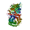
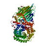
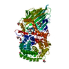
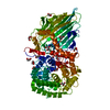
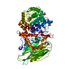
| Title | Crystal structure of TxGH116 from Thermoanaerobacterium xylanolyticum with isofagomine | ||||||||||||
 Components Components | Glucosylceramidase | ||||||||||||
 Keywords Keywords | HYDROLASE / TxGH116 / beta-glucosidase / acid/base mutant / Thermoanaerobacterium xylanolyticum / cellobiose | ||||||||||||
| Function / homology |  Function and homology information Function and homology informationglucosylceramidase / glucosylceramide catabolic process / glucosylceramidase activity / beta-glucosidase activity / carbohydrate metabolic process / membrane / metal ion binding Similarity search - Function | ||||||||||||
| Biological species |  Thermoanaerobacterium xylanolyticum LX-11 (bacteria) Thermoanaerobacterium xylanolyticum LX-11 (bacteria) | ||||||||||||
| Method |  X-RAY DIFFRACTION / SYNCHROTRON / MOLECULAR REPLACEMENT / Resolution: 2 Å X-RAY DIFFRACTION / SYNCHROTRON / MOLECULAR REPLACEMENT / Resolution: 2 Å | ||||||||||||
 Authors Authors | Pengthaisong, S. / Ketudat Cairns, J.R. | ||||||||||||
| Funding support |  Thailand, 3items Thailand, 3items
| ||||||||||||
 Citation Citation | Journal: Chem.Biol.Interact. / Year: 2023 Title: Structural basis for inhibition of a GH116 beta-glucosidase and its missense mutants by GBA2 inhibitors: Crystallographic and quantum chemical study. Authors: Meelua, W. / Thinkumrob, N. / Saparpakorn, P. / Pengthaisong, S. / Hannongbua, S. / Ketudat Cairns, J.R. / Jitonnom, J. | ||||||||||||
| History |
|
- Structure visualization
| Structure viewer | Molecule: MolmilJmol/JSmol |
|---|
- Downloads & links
-Download
| PDBx/mmCIF format | 8jbo.cif.gz | 345.6 KB | Display | PDBx/mmCIF format |
|---|---|---|---|---|
| PDB format | pdb8jbo.ent.gz | 274.6 KB | Display | PDB format |
| PDBx/mmJSON format | 8jbo.json.gz | Tree view | PDBx/mmJSON format | |
| Others |  Other downloads Other downloads |
-Validation report
| Arichive directory | https://data.pdbj.org/pub/pdb/validation_reports/jb/8jboftp://data.pdbj.org/pub/pdb/validation_reports/jb/8jbo | HTTPS FTP |
|---|
-Related structure data
| Related structure data | |
|---|---|
| Similar structure data |
-Links
 PDBj
PDBj


- Assembly
| Deposited unit | 
| ||||||||||||||||||||||||||||||
|---|---|---|---|---|---|---|---|---|---|---|---|---|---|---|---|---|---|---|---|---|---|---|---|---|---|---|---|---|---|---|---|
| 1 | 
| ||||||||||||||||||||||||||||||
| 2 | 
| ||||||||||||||||||||||||||||||
| Unit cell |
| ||||||||||||||||||||||||||||||
| Noncrystallographic symmetry (NCS) | NCS domain:
NCS domain segments: Component-ID: 1 / Ens-ID: 1 / Beg auth comp-ID: ASP / Beg label comp-ID: ASP / End auth comp-ID: VAL / End label comp-ID: VAL / Refine code: 1 / Auth seq-ID: 31 - 801 / Label seq-ID: 15 - 785
NCS oper:
|
-Components
-Protein , 1 types, 2 molecules AB
| #1: Protein | Mass: 91723.109 Da / Num. of mol.: 2 Source method: isolated from a genetically manipulated source Source: (gene. exp.) Thermoanaerobacterium xylanolyticum LX-11 (bacteria)Strain: LX-11 / Gene: Thexy_2211 / Plasmid: pET30a / Production host: |
|---|
-Non-polymers , 5 types, 994 molecules 


| #2: Chemical |  Mass: 147.172 Da / Num. of mol.: 2 / Source method: obtained synthetically / Formula: C6H13NO3 / Feature type: SUBJECT OF INVESTIGATION Mass: 147.172 Da / Num. of mol.: 2 / Source method: obtained synthetically / Formula: C6H13NO3 / Feature type: SUBJECT OF INVESTIGATION#3: Chemical |  Mass: 40.078 Da / Num. of mol.: 3 / Source method: obtained synthetically / Formula: Ca / Feature type: SUBJECT OF INVESTIGATION Mass: 40.078 Da / Num. of mol.: 3 / Source method: obtained synthetically / Formula: Ca / Feature type: SUBJECT OF INVESTIGATION#4: Chemical | ChemComp-GOL /  Mass: 92.094 Da / Num. of mol.: 20 / Source method: obtained synthetically / Formula: C3H8O3 / Feature type: SUBJECT OF INVESTIGATION Mass: 92.094 Da / Num. of mol.: 20 / Source method: obtained synthetically / Formula: C3H8O3 / Feature type: SUBJECT OF INVESTIGATION#5: Chemical |  Mass: 62.068 Da / Num. of mol.: 2 / Source method: obtained synthetically / Formula: C2H6O2 / Feature type: SUBJECT OF INVESTIGATION Mass: 62.068 Da / Num. of mol.: 2 / Source method: obtained synthetically / Formula: C2H6O2 / Feature type: SUBJECT OF INVESTIGATION#6: Water | ChemComp-HOH / | Mass: 18.015 Da / Num. of mol.: 967 / Source method: isolated from a natural source / Formula: H2O |
|---|
-Details
| Has ligand of interest | Y |
|---|
-Experimental details
-Experiment
| Experiment | Method: X-RAY DIFFRACTION / Number of used crystals: 1 |
|---|
- Sample preparation
| Crystal | Density Matthews: 2.2 Å3/Da / Density % sol: 47.7 % |
|---|---|
| Crystal grow | Temperature: 288 K / Method: vapor diffusion, hanging drop / pH: 5.5 Details: 0.2 M AMMONIUM SULFATE, 23% PEG 3000, 0.1 M MES, PH 5.5 |
-Data collection
| Diffraction | Mean temperature: 100 K / Serial crystal experiment: N |
|---|---|
| Diffraction source | Source: SYNCHROTRON / Site: SPring-8  / Beamline: BL44XU / Wavelength: 0.9 Å / Beamline: BL44XU / Wavelength: 0.9 Å |
| Detector | Type: RAYONIX MX300HE / Detector: CCD / Date: Nov 12, 2015 |
| Radiation | Protocol: SINGLE WAVELENGTH / Monochromatic (M) / Laue (L): M / Scattering type: x-ray |
| Radiation wavelength | Wavelength: 0.9 Å / Relative weight: 1 |
| Reflection | Resolution: 2→50.01 Å / Num. obs: 109281 / % possible obs: 98.8 % / Redundancy: 6.4 % / Rmerge(I) obs: 0.114 / Net I/σ(I): 26.7 |
| Reflection shell | Resolution: 2→2.03 Å / Redundancy: 5.8 % / Rmerge(I) obs: 0.447 / Mean I/σ(I) obs: 5.2 / Num. unique obs: 5192 / CC1/2: 0.884 / % possible all: 94.5 |
- Processing
| Software |
| ||||||||||||||||||||||||||||||||||||||||||||||||||||||||||||
|---|---|---|---|---|---|---|---|---|---|---|---|---|---|---|---|---|---|---|---|---|---|---|---|---|---|---|---|---|---|---|---|---|---|---|---|---|---|---|---|---|---|---|---|---|---|---|---|---|---|---|---|---|---|---|---|---|---|---|---|---|---|
| Refinement | Method to determine structure: MOLECULAR REPLACEMENT / Resolution: 2→50.01 Å / Cor.coef. Fo:Fc: 0.965 / Cor.coef. Fo:Fc free: 0.945 / SU B: 3.592 / SU ML: 0.098 / Cross valid method: THROUGHOUT / σ(F): 0 / ESU R: 0.166 / ESU R Free: 0.143 / Stereochemistry target values: MAXIMUM LIKELIHOOD Details: HYDROGENS HAVE BEEN ADDED IN THE RIDING POSITIONS U VALUES : REFINED INDIVIDUALLY
| ||||||||||||||||||||||||||||||||||||||||||||||||||||||||||||
| Solvent computation | Ion probe radii: 0.8 Å / Shrinkage radii: 0.8 Å / VDW probe radii: 1.2 Å / Solvent model: MASK | ||||||||||||||||||||||||||||||||||||||||||||||||||||||||||||
| Displacement parameters | Biso max: 87.52 Å2 / Biso mean: 22.149 Å2 / Biso min: 8.84 Å2
| ||||||||||||||||||||||||||||||||||||||||||||||||||||||||||||
| Refinement step | Cycle: final / Resolution: 2→50.01 Å
| ||||||||||||||||||||||||||||||||||||||||||||||||||||||||||||
| Refine LS restraints |
| ||||||||||||||||||||||||||||||||||||||||||||||||||||||||||||
| Refine LS restraints NCS | Number: 12071 / Type: TIGHT THERMAL / Rms dev position: 2 Å / Weight position: 0.5 | ||||||||||||||||||||||||||||||||||||||||||||||||||||||||||||
| LS refinement shell | Resolution: 2→2.05 Å / Rfactor Rfree error: 0
|
