glucosylceramidase / glucosylceramide catabolic process / glucosylceramidase activity / beta-glucosidase activity / carbohydrate metabolic process / membrane / metal ion binding Similarity search - Function
Glycosyl-hydrolase family 116, catalytic region / Beta-glucosidase GBA2-type / Glycosyl-hydrolase family 116, N-terminal / : / Glycosyl-hydrolase family 116, catalytic region / beta-glucosidase 2, glycosyl-hydrolase family 116 N-term / Six-hairpin glycosidase-like superfamily / Six-hairpin glycosidase superfamily / Prokaryotic membrane lipoprotein lipid attachment site profile. Similarity search - Domain/homology
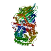
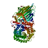
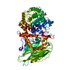
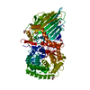
Mass: 91794.180 Da / Num. of mol.: 1 / Fragment: UNP residues 19-806 Source method: isolated from a genetically manipulated source Details: The author believe that the E.C. number and the enzyme name in UniProt F6BL85 is incorrect. Source: (gene. exp.) Thermoanaerobacterium xylanolyticum LX-11 (bacteria) Strain: LX-11 / Gene: Thexy_2211 / Production host: Escherichia coli BL21(DE3) (bacteria) / Strain (production host): BL21(DE3) / References: UniProt: F6BL85, beta-glucosidase
Protocol: SINGLE WAVELENGTH / Monochromatic (M) / Laue (L): M / Scattering type: x-ray
Radiation wavelength
Wavelength: 0.9 Å / Relative weight: 1
Reflection
Resolution: 1.61→50 Å / Num. obs: 102998 / % possible obs: 99 % / Redundancy: 3.5 % / Rmerge(I) obs: 0.05 / Net I/σ(I): 34.4
Reflection shell
Resolution: 1.61→1.64 Å / Redundancy: 3.5 % / Rmerge(I) obs: 0.322 / Mean I/σ(I) obs: 5.9 / % possible all: 99.2
-
Processing
Software
Name
Version
Classification
REFMAC
5.8.0073
refinement
HKL-2000
datareduction
HKL-2000
datascaling
PHENIX
phasing
Refinement
Method to determine structure: SAD / Resolution: 1.61→50 Å / Cor.coef. Fo:Fc: 0.972 / Cor.coef. Fo:Fc free: 0.965 / SU B: 1.393 / SU ML: 0.049 / Cross valid method: THROUGHOUT / ESU R: 0.08 / ESU R Free: 0.077 / Stereochemistry target values: MAXIMUM LIKELIHOOD / Details: HYDROGENS HAVE BEEN ADDED IN THE RIDING POSITIONS
Rfactor
Num. reflection
% reflection
Selection details
Rfree
0.17532
5069
4.9 %
RANDOM
Rwork
0.15378
-
-
-
obs
0.15482
97906
98.33 %
-
Solvent computation
Ion probe radii: 0.8 Å / Shrinkage radii: 0.8 Å / VDW probe radii: 1.2 Å / Solvent model: MASK
 Movie
Movie Controller

 Yorodumi
 Open data
 Basic information
 Components
 Keywords
 Function and homology information
 Thermoanaerobacterium xylanolyticum LX-11 (bacteria)
 X-RAY DIFFRACTION /
  Authors
 Thailand,
  Japan, 3items
  Citation
 Structure visualization
 Downloads & links
 Other downloads
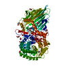


 PDBj
PDBj


 Assembly
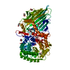

 Mass: 92.094 Da / Num. of mol.: 16 / Source method: obtained synthetically / Formula: C3H8O3
 Mass: 40.078 Da / Num. of mol.: 1 / Source method: obtained synthetically / Formula: Ca
 Mass: 62.068 Da / Num. of mol.: 1 / Source method: obtained synthetically / Formula: C2H6O2
 Mass: 18.015 Da / Num. of mol.: 583 / Source method: isolated from a natural source / Formula: H2O
 Sample preparation
 Processing