 Movie
Movie Controller

[English] 日本語
 Yorodumi
- PDB-8j4v: Structure of Mycobacterium thermoresistibile NrdI(oxidised) deter... -
+ Open data

- Basic information
| Entry | Database: PDB / ID: 8j4v | ||||||
|---|---|---|---|---|---|---|---|
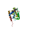
| Title | Structure of Mycobacterium thermoresistibile NrdI(oxidised) determined at 1.1 angstrom resolution | ||||||
 Components Components | Protein NrdI | ||||||
 Keywords Keywords | FLAVOPROTEIN / Ribonucleotide reductase accessory protein. Metal cofactor assembly | ||||||
| Function / homology | Ribonucleotide reductase, class Ib, NrdI, bacterial / Ribonucleotide reductase Class Ib, NrdI / NrdI Flavodoxin like / Flavoprotein-like superfamily / FMN binding / FLAVIN MONONUCLEOTIDE / PHOSPHATE ION / Protein NrdI Function and homology information Function and homology information | ||||||
| Biological species |  Mycolicibacterium thermoresistibile ATCC 19527 (bacteria) Mycolicibacterium thermoresistibile ATCC 19527 (bacteria) | ||||||
| Method |  X-RAY DIFFRACTION / SYNCHROTRON / MOLECULAR REPLACEMENT / Resolution: 1.11 Å X-RAY DIFFRACTION / SYNCHROTRON / MOLECULAR REPLACEMENT / Resolution: 1.11 Å | ||||||
 Authors Authors | Yadav, L.R. / Mande, S.C. | ||||||
| Funding support |  India, 1items India, 1items
| ||||||
 Citation Citation | Journal: Curr Res Struct Biol / Year: 2024 Title: Structural insights into the initiation of free radical formation in the Class Ib ribonucleotide reductases in Mycobacteria. Authors: Yadav, L.R. / Sharma, V. / Shanmugam, M. / Mande, S.C. | ||||||
| History |
|
- Structure visualization
| Structure viewer | Molecule: MolmilJmol/JSmol |
|---|
- Downloads & links
-Download
| PDBx/mmCIF format | 8j4v.cif.gz | 96.7 KB | Display | PDBx/mmCIF format |
|---|---|---|---|---|
| PDB format | pdb8j4v.ent.gz | 59.8 KB | Display | PDB format |
| PDBx/mmJSON format | 8j4v.json.gz | Tree view | PDBx/mmJSON format | |
| Others |  Other downloads Other downloads |
-Validation report
| Arichive directory | https://data.pdbj.org/pub/pdb/validation_reports/j4/8j4vftp://data.pdbj.org/pub/pdb/validation_reports/j4/8j4v | HTTPS FTP |
|---|
-Related structure data

-Links
 PDBj
PDBj- Assembly
| Deposited unit | 
| ||||||||||||
|---|---|---|---|---|---|---|---|---|---|---|---|---|---|
| 1 |
| ||||||||||||
| Unit cell |
|
-Components
| #1: Protein | Mass: 16442.625 Da / Num. of mol.: 1 Source method: isolated from a genetically manipulated source Details: Ribonucleotide reductase Metal cofactor Source: (gene. exp.) Mycolicibacterium thermoresistibile ATCC 19527 (bacteria)Strain: ATCC 19527 / Gene: nrdI / Plasmid: pET32a+ / Production host: |
|---|---|
| #2: Chemical | ChemComp-FMN /   Mass: 456.344 Da / Num. of mol.: 1 / Source method: obtained synthetically / Formula: C17H21N4O9P / Feature type: SUBJECT OF INVESTIGATION Mass: 456.344 Da / Num. of mol.: 1 / Source method: obtained synthetically / Formula: C17H21N4O9P / Feature type: SUBJECT OF INVESTIGATION |
| #3: Chemical | ChemComp-PO4 /   Mass: 94.971 Da / Num. of mol.: 1 / Source method: obtained synthetically / Formula: PO4 Mass: 94.971 Da / Num. of mol.: 1 / Source method: obtained synthetically / Formula: PO4 |
| #4: Chemical | ChemComp-GOL /   Mass: 92.094 Da / Num. of mol.: 1 / Source method: obtained synthetically / Formula: C3H8O3 Mass: 92.094 Da / Num. of mol.: 1 / Source method: obtained synthetically / Formula: C3H8O3 |
| #5: Water | ChemComp-HOH /  Mass: 18.015 Da / Num. of mol.: 127 / Source method: isolated from a natural source / Formula: H2O Mass: 18.015 Da / Num. of mol.: 127 / Source method: isolated from a natural source / Formula: H2O |
| Has ligand of interest | Y |
| Has protein modification | N |
-Experimental details
-Experiment
| Experiment | Method: X-RAY DIFFRACTION / Number of used crystals: 1 |
|---|
- Sample preparation
| Crystal | Density Matthews: 1.83 Å3/Da / Density % sol: 32.87 % Description: Rectangular yellow color small sized crystals of 50-80 micron |
|---|---|
| Crystal grow | Temperature: 293.15 K / Method: vapor diffusion, sitting drop / pH: 7.8 Details: 0.15M potassium phosphate monobasic and 18% PEG 3350 Temp details: 293.15 |
-Data collection
| Diffraction | Mean temperature: 100 K / Ambient temp details: 100 / Serial crystal experiment: N |
|---|---|
| Diffraction source | Source: SYNCHROTRON / Site: Diamond  / Beamline: I04 / Wavelength: 0.9795 Å / Beamline: I04 / Wavelength: 0.9795 Å |
| Detector | Type: DECTRIS PILATUS 6M / Detector: PIXEL / Date: Dec 11, 2017 |
| Radiation | Protocol: SINGLE WAVELENGTH / Monochromatic (M) / Laue (L): M / Scattering type: x-ray |
| Radiation wavelength | Wavelength: 0.9795 Å / Relative weight: 1 |
| Reflection | Resolution: 1.11→44.98 Å / Num. obs: 47159 / % possible obs: 99.77 % / Redundancy: 12.6 % / Biso Wilson estimate: 7.59 Å2 / CC1/2: 0.793 / CC star: 0.94 / Net I/σ(I): 35.82 |
| Reflection shell | Resolution: 1.11→1.15 Å / Redundancy: 11.2 % / Num. unique obs: 4696 / CC1/2: 0.628 / CC star: 0.878 / % possible all: 99.68 |
- Processing
| Software |
| ||||||||||||||||||||||||||||||||||||||||||||||||||||||||||||||||||||||||||||||||||||||||||||||||||||||||||||||||||||||||||||||
|---|---|---|---|---|---|---|---|---|---|---|---|---|---|---|---|---|---|---|---|---|---|---|---|---|---|---|---|---|---|---|---|---|---|---|---|---|---|---|---|---|---|---|---|---|---|---|---|---|---|---|---|---|---|---|---|---|---|---|---|---|---|---|---|---|---|---|---|---|---|---|---|---|---|---|---|---|---|---|---|---|---|---|---|---|---|---|---|---|---|---|---|---|---|---|---|---|---|---|---|---|---|---|---|---|---|---|---|---|---|---|---|---|---|---|---|---|---|---|---|---|---|---|---|---|---|---|---|
| Refinement | Method to determine structure: MOLECULAR REPLACEMENT / Resolution: 1.11→44.98 Å / SU ML: 0.0884 / Cross valid method: FREE R-VALUE / σ(F): 1.33 / Phase error: 14.1213 Stereochemistry target values: GeoStd + Monomer Library + CDL v1.2
| ||||||||||||||||||||||||||||||||||||||||||||||||||||||||||||||||||||||||||||||||||||||||||||||||||||||||||||||||||||||||||||||
| Solvent computation | Shrinkage radii: 0.9 Å / VDW probe radii: 1.11 Å / Solvent model: FLAT BULK SOLVENT MODEL | ||||||||||||||||||||||||||||||||||||||||||||||||||||||||||||||||||||||||||||||||||||||||||||||||||||||||||||||||||||||||||||||
| Displacement parameters | Biso mean: 13.49 Å2 | ||||||||||||||||||||||||||||||||||||||||||||||||||||||||||||||||||||||||||||||||||||||||||||||||||||||||||||||||||||||||||||||
| Refinement step | Cycle: LAST / Resolution: 1.11→44.98 Å
| ||||||||||||||||||||||||||||||||||||||||||||||||||||||||||||||||||||||||||||||||||||||||||||||||||||||||||||||||||||||||||||||
| Refine LS restraints |
| ||||||||||||||||||||||||||||||||||||||||||||||||||||||||||||||||||||||||||||||||||||||||||||||||||||||||||||||||||||||||||||||
| LS refinement shell |
|
