 Movie
Movie Controller

[English] 日本語
 Yorodumi
- PDB-8ix3: Cryo-EM structure of SARS-CoV-2 BA.4/5 spike protein in complex w... -
+ Open data

- Basic information
| Entry | Database: PDB / ID: 8ix3 | ||||||
|---|---|---|---|---|---|---|---|
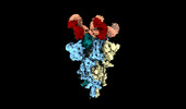
| Title | Cryo-EM structure of SARS-CoV-2 BA.4/5 spike protein in complex with 1G11 (local refinement) | ||||||
 Components Components |
| ||||||
 Keywords Keywords | VIRAL PROTEIN/IMMUNE SYSTEM / SARS-CoV-2 / Neutralizing antibody / Cryo-EM / VIRAL PROTEIN / VIRAL PROTEIN-IMMUNE SYSTEM complex | ||||||
| Function / homology |  Function and homology information Function and homology informationMaturation of spike protein / viral translation / Translation of Structural Proteins / Virion Assembly and Release / host cell surface / host extracellular space / suppression by virus of host tetherin activity / Induction of Cell-Cell Fusion / structural constituent of virion / entry receptor-mediated virion attachment to host cell ...Maturation of spike protein / viral translation / Translation of Structural Proteins / Virion Assembly and Release / host cell surface / host extracellular space / suppression by virus of host tetherin activity / Induction of Cell-Cell Fusion / structural constituent of virion / entry receptor-mediated virion attachment to host cell / host cell endoplasmic reticulum-Golgi intermediate compartment membrane / receptor-mediated endocytosis of virus by host cell / membrane fusion / Attachment and Entry / positive regulation of viral entry into host cell / receptor-mediated virion attachment to host cell / receptor ligand activity / host cell surface receptor binding / fusion of virus membrane with host plasma membrane / fusion of virus membrane with host endosome membrane / viral envelope / virion attachment to host cell / SARS-CoV-2 activates/modulates innate and adaptive immune responses / host cell plasma membrane / virion membrane / identical protein binding / membrane / plasma membrane Similarity search - Function | ||||||
| Biological species |  Homo sapiens (human) Homo sapiens (human)  Severe acute respiratory syndrome coronavirus 2 Severe acute respiratory syndrome coronavirus 2 | ||||||
| Method | ELECTRON MICROSCOPY / single particle reconstruction / cryo EM / Resolution: 3.98 Å | ||||||
 Authors Authors | Sun, H. / Jiang, Y. / Zheng, Z. / Zheng, Q. / Li, S. | ||||||
| Funding support | 1items
| ||||||
 Citation Citation | Journal: J Virol / Year: 2023 Title: Structural basis for broad neutralization of human antibody against Omicron sublineages and evasion by XBB variant. Authors: Hui Sun / Yizhen Wang / Xiuting Chen / Yanan Jiang / Siling Wang / Yang Huang / Liqin Liu / Yu Li / Miaolin Lan / Huilin Guo / Quan Yuan / Yali Zhang / Tingting Li / Hai Yu / Ying Gu / Jun ...Authors: Hui Sun / Yizhen Wang / Xiuting Chen / Yanan Jiang / Siling Wang / Yang Huang / Liqin Liu / Yu Li / Miaolin Lan / Huilin Guo / Quan Yuan / Yali Zhang / Tingting Li / Hai Yu / Ying Gu / Jun Zhang / Shaowei Li / Zizheng Zheng / Qingbing Zheng / Ningshao Xia /  Abstract: The ongoing COVID-19 pandemic has been characterized by the emergence of new SARS-CoV-2 variants including the highly transmissible Omicron XBB sublineages, which have shown significant resistance to ...The ongoing COVID-19 pandemic has been characterized by the emergence of new SARS-CoV-2 variants including the highly transmissible Omicron XBB sublineages, which have shown significant resistance to neutralizing antibodies (nAbs). This resistance has led to decreased vaccine effectiveness and therefore result in breakthrough infections and reinfections, which continuously threaten public health. To date, almost all available therapeutic nAbs, including those authorized under Emergency Use Authorization nAbs that were previously clinically useful against early strains, have recently been found to be ineffective against newly emerging variants. In this study, we provide a comprehensive structural basis about how the Class 3 nAbs, including 1G11 in this study and noted LY-CoV1404, are evaded by the newly emerged SARS-CoV-2 variants. | ||||||
| History |
|
- Structure visualization
| Structure viewer | Molecule: MolmilJmol/JSmol |
|---|
- Downloads & links
-Download
| PDBx/mmCIF format | 8ix3.cif.gz | 87.1 KB | Display | PDBx/mmCIF format |
|---|---|---|---|---|
| PDB format | pdb8ix3.ent.gz | 64.5 KB | Display | PDB format |
| PDBx/mmJSON format | 8ix3.json.gz | Tree view | PDBx/mmJSON format | |
| Others |  Other downloads Other downloads |
-Validation report
| Summary document | 8ix3_validation.pdf.gz | 1.3 MB | Display | wwPDB validaton report |
|---|---|---|---|---|
| Full document | 8ix3_full_validation.pdf.gz | 1.3 MB | Display | |
| Data in XML | 8ix3_validation.xml.gz | 32.1 KB | Display | |
| Data in CIF | 8ix3_validation.cif.gz | 42.6 KB | Display | |
| Arichive directory | https://data.pdbj.org/pub/pdb/validation_reports/ix/8ix3ftp://data.pdbj.org/pub/pdb/validation_reports/ix/8ix3 | HTTPS FTP |
-Related structure data
| Related structure data |  35788MC M: map data used to model this data C: citing same article ( |
|---|---|
| Similar structure data |

-Links
 PDBj
PDBj


- Assembly
| Deposited unit | 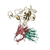
|
|---|---|
| 1 |
|
-Components
| #1: Antibody | Mass: 11628.971 Da / Num. of mol.: 1 Source method: isolated from a genetically manipulated source Source: (gene. exp.) Homo sapiens (human) / Production host:   Cricetulus griseus (Chinese hamster) Cricetulus griseus (Chinese hamster) |
|---|---|
| #2: Antibody | Mass: 13772.198 Da / Num. of mol.: 1 Source method: isolated from a genetically manipulated source Source: (gene. exp.) Homo sapiens (human) / Production host: Cricetulus griseus (Chinese hamster) |
| #3: Protein | Mass: 23949.986 Da / Num. of mol.: 1 Source method: isolated from a genetically manipulated source Source: (gene. exp.) Severe acute respiratory syndrome coronavirus 2Gene: S, 2 / Cell line (production host): HEK293 / Production host: Homo sapiens (human) / References: UniProt: P0DTC2 |
-Experimental details
-Experiment
| Experiment | Method: ELECTRON MICROSCOPY |
|---|---|
| EM experiment | Aggregation state: PARTICLE / 3D reconstruction method: single particle reconstruction |
- Sample preparation
| Component |
| ||||||||||||||||||||||||
|---|---|---|---|---|---|---|---|---|---|---|---|---|---|---|---|---|---|---|---|---|---|---|---|---|---|
| Source (natural) |
| ||||||||||||||||||||||||
| Source (recombinant) |
| ||||||||||||||||||||||||
| Buffer solution | pH: 7.4 | ||||||||||||||||||||||||
| Specimen | Embedding applied: NO / Shadowing applied: NO / Staining applied: NO / Vitrification applied: YES | ||||||||||||||||||||||||
| Vitrification | Cryogen name: ETHANE |
- Electron microscopy imaging
| Experimental equipment |  Model: Tecnai F30 / Image courtesy: FEI Company |
|---|---|
| Microscopy | Model: FEI TECNAI F30 |
| Electron gun | Electron source:  FIELD EMISSION GUN / Accelerating voltage: 300 kV / Illumination mode: FLOOD BEAM FIELD EMISSION GUN / Accelerating voltage: 300 kV / Illumination mode: FLOOD BEAM |
| Electron lens | Mode: BRIGHT FIELD / Nominal defocus max: 2000 nm / Nominal defocus min: 800 nm |
| Image recording | Electron dose: 60 e/Å2 / Film or detector model: GATAN K3 (6k x 4k) |
- Processing
| CTF correction | Type: PHASE FLIPPING AND AMPLITUDE CORRECTION |
|---|---|
| 3D reconstruction | Resolution: 3.98 Å / Resolution method: FSC 0.143 CUT-OFF / Num. of particles: 275583 / Symmetry type: POINT |
