 Movie
Movie Controller

[English] 日本語
 Yorodumi
- PDB-8itm: Cryo-EM structure of GIPR splice variant 2 (SV2) in complex with ... -
+ Open data

- Basic information
| Entry | Database: PDB / ID: 8itm | |||||||||||||||||||||||||||||||||
|---|---|---|---|---|---|---|---|---|---|---|---|---|---|---|---|---|---|---|---|---|---|---|---|---|---|---|---|---|---|---|---|---|---|---|
| Title | Cryo-EM structure of GIPR splice variant 2 (SV2) in complex with Gs protein | |||||||||||||||||||||||||||||||||
 Components Components |
| |||||||||||||||||||||||||||||||||
 Keywords Keywords | STRUCTURAL PROTEIN / Cryo-electron microscopy / G protein-coupled receptor / splice variant / receptor activation. | |||||||||||||||||||||||||||||||||
| Function / homology |  Function and homology information Function and homology informationgastric inhibitory peptide receptor activity / glucagon family peptide binding / gastric inhibitory peptide signaling pathway / endocrine pancreas development / response to fatty acid / sensory perception of chemical stimulus / desensitization of G protein-coupled receptor signaling pathway / mu-type opioid receptor binding / corticotropin-releasing hormone receptor 1 binding / G-protein activation ...gastric inhibitory peptide receptor activity / glucagon family peptide binding / gastric inhibitory peptide signaling pathway / endocrine pancreas development / response to fatty acid / sensory perception of chemical stimulus / desensitization of G protein-coupled receptor signaling pathway / mu-type opioid receptor binding / corticotropin-releasing hormone receptor 1 binding / G-protein activation / Activation of the phototransduction cascade / Glucagon-type ligand receptors / Thromboxane signalling through TP receptor / Sensory perception of sweet, bitter, and umami (glutamate) taste / G beta:gamma signalling through PI3Kgamma / G beta:gamma signalling through CDC42 / Cooperation of PDCL (PhLP1) and TRiC/CCT in G-protein beta folding / Activation of G protein gated Potassium channels / Inhibition of voltage gated Ca2+ channels via Gbeta/gamma subunits / Ca2+ pathway / G alpha (z) signalling events / G protein-coupled peptide receptor activity / High laminar flow shear stress activates signaling by PIEZO1 and PECAM1:CDH5:KDR in endothelial cells / Glucagon-like Peptide-1 (GLP1) regulates insulin secretion / Vasopressin regulates renal water homeostasis via Aquaporins / Adrenaline,noradrenaline inhibits insulin secretion / ADP signalling through P2Y purinoceptor 12 / G alpha (q) signalling events / beta-2 adrenergic receptor binding / G alpha (i) signalling events / Thrombin signalling through proteinase activated receptors (PARs) / Activation of G protein gated Potassium channels / G-protein activation / G beta:gamma signalling through PI3Kgamma / Prostacyclin signalling through prostacyclin receptor / G beta:gamma signalling through PLC beta / ADP signalling through P2Y purinoceptor 1 / Thromboxane signalling through TP receptor / Presynaptic function of Kainate receptors / G beta:gamma signalling through CDC42 / Inhibition of voltage gated Ca2+ channels via Gbeta/gamma subunits / G alpha (12/13) signalling events / Glucagon-type ligand receptors / G beta:gamma signalling through BTK / ADP signalling through P2Y purinoceptor 12 / Adrenaline,noradrenaline inhibits insulin secretion / Cooperation of PDCL (PhLP1) and TRiC/CCT in G-protein beta folding / Ca2+ pathway / G alpha (z) signalling events / Thrombin signalling through proteinase activated receptors (PARs) / Extra-nuclear estrogen signaling / G alpha (q) signalling events / G alpha (s) signalling events / photoreceptor outer segment membrane / spectrin binding / Glucagon-like Peptide-1 (GLP1) regulates insulin secretion / G alpha (i) signalling events / High laminar flow shear stress activates signaling by PIEZO1 and PECAM1:CDH5:KDR in endothelial cells / Vasopressin regulates renal water homeostasis via Aquaporins / alkylglycerophosphoethanolamine phosphodiesterase activity / sensory perception of taste / regulation of insulin secretion / peptide hormone binding / response to axon injury / response to glucose / photoreceptor outer segment / D1 dopamine receptor binding / retina development in camera-type eye / activation of adenylate cyclase activity / adenylate cyclase-activating adrenergic receptor signaling pathway / insulin-like growth factor receptor binding / cardiac muscle cell apoptotic process / photoreceptor inner segment / response to nutrient / ionotropic glutamate receptor binding / adenylate cyclase activator activity / generation of precursor metabolites and energy / response to calcium ion / cell population proliferation / positive regulation of insulin secretion / G-protein beta/gamma-subunit complex binding / adenylate cyclase-modulating G protein-coupled receptor signaling pathway / phospholipase C-activating G protein-coupled receptor signaling pathway / Glucagon-type ligand receptors / transmembrane signaling receptor activity / cellular response to catecholamine stimulus / adenylate cyclase-activating dopamine receptor signaling pathway / cellular response to prostaglandin E stimulus / heterotrimeric G-protein complex / G-protein beta-subunit binding / positive regulation of cytosolic calcium ion concentration / signaling receptor complex adaptor activity / adenylate cyclase-activating G protein-coupled receptor signaling pathway / cell body / GTPase binding / cellular response to hypoxia / G alpha (s) signalling events / Hydrolases; Acting on acid anhydrides; Acting on GTP to facilitate cellular and subcellular movement / cell surface receptor signaling pathway / G protein-coupled receptor signaling pathway Similarity search - Function | |||||||||||||||||||||||||||||||||
| Biological species |  Homo sapiens (human) Homo sapiens (human) synthetic construct (others) | |||||||||||||||||||||||||||||||||
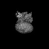
| Method | ELECTRON MICROSCOPY / single particle reconstruction / cryo EM / Resolution: 3.13 Å | |||||||||||||||||||||||||||||||||
 Authors Authors | Zhao, F.H. / Hang, K.N. / Zhou, Q.T. / Shao, L.J. / Li, H. / Li, W.Z. / Lin, S. / Dai, A.T. / Cai, X.Q. / Liu, Y.Y. ...Zhao, F.H. / Hang, K.N. / Zhou, Q.T. / Shao, L.J. / Li, H. / Li, W.Z. / Lin, S. / Dai, A.T. / Cai, X.Q. / Liu, Y.Y. / Xu, Y.N. / Feng, W.B. / Yang, D.H. / Wang, M.W. | |||||||||||||||||||||||||||||||||
| Funding support |  China, 6items China, 6items
| |||||||||||||||||||||||||||||||||
 Citation Citation | Journal: Proc Natl Acad Sci U S A / Year: 2023 Title: Molecular basis of signal transduction mediated by the human GIPR splice variants. Authors: Fenghui Zhao / Kaini Hang / Qingtong Zhou / Lijun Shao / Hao Li / Wenzhuo Li / Shi Lin / Antao Dai / Xiaoqing Cai / Yanyun Liu / Yingna Xu / Wenbo Feng / Dehua Yang / Ming-Wei Wang /  Abstract: Glucose-dependent insulinotropic polypeptide receptor (GIPR) is a potential drug target for metabolic disorders. It works with glucagon-like peptide-1 receptor and glucagon receptor in humans to ...Glucose-dependent insulinotropic polypeptide receptor (GIPR) is a potential drug target for metabolic disorders. It works with glucagon-like peptide-1 receptor and glucagon receptor in humans to maintain glucose homeostasis. Unlike the other two receptors, GIPR has at least 13 reported splice variants (SVs), more than half of which have sequence variations at either C or N terminus. To explore their roles in endogenous peptide-mediated GIPR signaling, we determined the cryoelectron microscopy (cryo-EM) structures of the two N terminus-altered SVs (referred as GIPR-202 and GIPR-209 in the Ensembl database, SV1 and SV2 here, respectively) and investigated the outcome of coexpressing each of them in question with GIPR in HEK293T cells with respect to ligand binding, receptor expression, cAMP (adenosine 3,5-cyclic monophosphate) accumulation, β-arrestin recruitment, and cell surface localization. It was found that while both N terminus-altered SVs of GIPR neither bound to the hormone nor elicited signal transduction per se, they suppressed ligand binding and cAMP accumulation of GIPR. Meanwhile, SV1 reduced GIPR-mediated β-arrestin 2 responses. The cryo-EM structures of SV1 and SV2 showed that they reorganized the extracellular halves of transmembrane helices 1, 6, and 7 and extracellular loops 2 and 3 to adopt a ligand-binding pocket-occupied conformation, thereby losing binding ability to the peptide. The results suggest a form of signal bias that is constitutive and ligand-independent, thus expanding our knowledge of biased signaling beyond pharmacological manipulation (i.e., ligand specific) as well as constitutive and ligand-independent (e.g., SV1 of the growth hormone-releasing hormone receptor). | |||||||||||||||||||||||||||||||||
| History |
|
- Structure visualization
| Structure viewer | Molecule: MolmilJmol/JSmol |
|---|
- Downloads & links
-Download
| PDBx/mmCIF format | 8itm.cif.gz | 196.3 KB | Display | PDBx/mmCIF format |
|---|---|---|---|---|
| PDB format | pdb8itm.ent.gz | 148.4 KB | Display | PDB format |
| PDBx/mmJSON format | 8itm.json.gz | Tree view | PDBx/mmJSON format | |
| Others |  Other downloads Other downloads |
-Validation report
| Arichive directory | https://data.pdbj.org/pub/pdb/validation_reports/it/8itmftp://data.pdbj.org/pub/pdb/validation_reports/it/8itm | HTTPS FTP |
|---|
-Related structure data
| Related structure data | 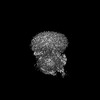 35707MC 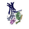 8itlC M: map data used to model this data C: citing same article ( |
|---|---|
| Similar structure data |
-Links
 PDBj
PDBj


- Assembly
| Deposited unit | 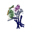
|
|---|---|
| 1 |
|
-Components
| #1: Protein | Mass: 41358.254 Da / Num. of mol.: 1 / Mutation: T284F Source method: isolated from a genetically manipulated source Source: (gene. exp.) Homo sapiens (human) / Gene: GIPR / Production host:   Spodoptera frugiperda (fall armyworm) / References: UniProt: P48546 Spodoptera frugiperda (fall armyworm) / References: UniProt: P48546 |
|---|---|
| #2: Protein | Mass: 45727.441 Da / Num. of mol.: 1 / Mutation: S54N,G226A,E268A,N271K,K274D,R280K,T284D,I285T Source method: isolated from a genetically manipulated source Source: (gene. exp.) Spodoptera frugiperda (fall armyworm) / References: UniProt: P04896 |
| #3: Protein | Mass: 40226.992 Da / Num. of mol.: 1 Source method: isolated from a genetically manipulated source Source: (gene. exp.) Spodoptera frugiperda (fall armyworm) / References: UniProt: P54311 |
| #4: Protein | Mass: 7861.143 Da / Num. of mol.: 1 Source method: isolated from a genetically manipulated source Source: (gene. exp.) Spodoptera frugiperda (fall armyworm) / References: UniProt: P63212 |
| #5: Antibody | Mass: 13885.439 Da / Num. of mol.: 1 Source method: isolated from a genetically manipulated source Source: (gene. exp.) synthetic construct (others) / Production host:  |
| Has protein modification | Y |
-Experimental details
-Experiment
| Experiment | Method: ELECTRON MICROSCOPY |
|---|---|
| EM experiment | Aggregation state: PARTICLE / 3D reconstruction method: single particle reconstruction |
- Sample preparation
| Component | Name: Cryo-EM structure of a splice variant of the GIPR(SV2) in complex with Gs protein Type: COMPLEX / Entity ID: all / Source: MULTIPLE SOURCES |
|---|---|
| Source (natural) | Organism: Homo sapiens (human) |
| Source (recombinant) | Organism: Spodoptera frugiperda (fall armyworm) |
| Buffer solution | pH: 7.4 |
| Specimen | Embedding applied: NO / Shadowing applied: NO / Staining applied: NO / Vitrification applied: YES |
| Vitrification | Cryogen name: ETHANE |
- Electron microscopy imaging
| Experimental equipment |  Model: Titan Krios / Image courtesy: FEI Company |
|---|---|
| Microscopy | Model: FEI TITAN KRIOS |
| Electron gun | Electron source: OTHER / Accelerating voltage: 300 kV / Illumination mode: OTHER |
| Electron lens | Mode: BRIGHT FIELD / Nominal defocus max: 2200 nm / Nominal defocus min: 1200 nm |
| Image recording | Electron dose: 80 e/Å2 / Film or detector model: GATAN K3 (6k x 4k) |
- Processing
| EM software | Name: PHENIX / Category: model refinement | ||||||||||||||||||||||||
|---|---|---|---|---|---|---|---|---|---|---|---|---|---|---|---|---|---|---|---|---|---|---|---|---|---|
| CTF correction | Type: PHASE FLIPPING AND AMPLITUDE CORRECTION | ||||||||||||||||||||||||
| 3D reconstruction | Resolution: 3.13 Å / Resolution method: DIFFRACTION PATTERN/LAYERLINES / Num. of particles: 463406 / Symmetry type: POINT | ||||||||||||||||||||||||
| Refine LS restraints |
|
