 Movie
Movie Controller

[English] 日本語
 Yorodumi
- PDB-8isq: Crystal structure of extended-spectrum class A beta-lactamase, CE... -
+ Open data

- Basic information
| Entry | Database: PDB / ID: 8isq | ||||||
|---|---|---|---|---|---|---|---|
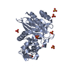
| Title | Crystal structure of extended-spectrum class A beta-lactamase, CESS-1 E166Q acylated by ampicillin | ||||||
 Components Components | Beta-lactamase | ||||||
 Keywords Keywords | HYDROLASE / extended-spectrum Class A beta-lactamase | ||||||
| Function / homology |  Function and homology information Function and homology informationbeta-lactam antibiotic catabolic process / beta-lactamase activity / beta-lactamase / response to antibiotic Similarity search - Function | ||||||
| Biological species |  Stenotrophomonas sp. KCTC 12332 (bacteria) Stenotrophomonas sp. KCTC 12332 (bacteria) | ||||||
| Method |  X-RAY DIFFRACTION / SYNCHROTRON / MOLECULAR REPLACEMENT / Resolution: 2.24 Å X-RAY DIFFRACTION / SYNCHROTRON / MOLECULAR REPLACEMENT / Resolution: 2.24 Å | ||||||
 Authors Authors | Jeong, B.G. / Kim, M.Y. / Jeong, C.S. / Do, H.W. / Lee, J.H. / Cha, S.S. | ||||||
| Funding support | 1items
| ||||||
 Citation Citation | Journal: Int J Antimicrob Agents / Year: 2024 Title: Characterization of the extended substrate spectrum of the class A beta-lactamase CESS-1 from Stenotrophomonas sp. and structure-based investigation into its substrate preference. Authors: Jeong, B.G. / Kim, M.Y. / Jeong, C.S. / Do, H. / Hwang, J. / Lee, J.H. / Cha, S.S. | ||||||
| History |
|
- Structure visualization
| Structure viewer | Molecule: MolmilJmol/JSmol |
|---|
- Downloads & links
-Download
| PDBx/mmCIF format | 8isq.cif.gz | 83 KB | Display | PDBx/mmCIF format |
|---|---|---|---|---|
| PDB format | pdb8isq.ent.gz | 47.9 KB | Display | PDB format |
| PDBx/mmJSON format | 8isq.json.gz | Tree view | PDBx/mmJSON format | |
| Others |  Other downloads Other downloads |
-Validation report
| Summary document | 8isq_validation.pdf.gz | 880.4 KB | Display | wwPDB validaton report |
|---|---|---|---|---|
| Full document | 8isq_full_validation.pdf.gz | 882.5 KB | Display | |
| Data in XML | 8isq_validation.xml.gz | 13.7 KB | Display | |
| Data in CIF | 8isq_validation.cif.gz | 19.2 KB | Display | |
| Arichive directory | https://data.pdbj.org/pub/pdb/validation_reports/is/8isqftp://data.pdbj.org/pub/pdb/validation_reports/is/8isq | HTTPS FTP |
-Related structure data
| Related structure data | 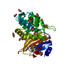 8isoSC 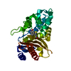 8ispC 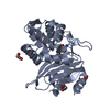 8isrC S: Starting model for refinement C: citing same article ( |
|---|---|
| Similar structure data |
-Links
 PDBj
PDBj


- Assembly
| Deposited unit | 
| ||||||||||||
|---|---|---|---|---|---|---|---|---|---|---|---|---|---|
| 1 |
| ||||||||||||
| Unit cell |
| ||||||||||||
| Components on special symmetry positions |
|
-Components
| #1: Protein | Mass: 28652.281 Da / Num. of mol.: 1 Source method: isolated from a genetically manipulated source Source: (gene. exp.) Stenotrophomonas sp. KCTC 12332 (bacteria)Gene: AXG53_04720 / Production host: |
|---|---|
| #2: Chemical | ChemComp-ZZ7 / (  Mass: 367.420 Da / Num. of mol.: 1 / Source method: obtained synthetically / Formula: C16H21N3O5S / Feature type: SUBJECT OF INVESTIGATION Mass: 367.420 Da / Num. of mol.: 1 / Source method: obtained synthetically / Formula: C16H21N3O5S / Feature type: SUBJECT OF INVESTIGATION |
| #3: Chemical | ChemComp-BTB /   Mass: 209.240 Da / Num. of mol.: 1 / Source method: obtained synthetically / Formula: C8H19NO5 / Comment: pH buffer*YM Mass: 209.240 Da / Num. of mol.: 1 / Source method: obtained synthetically / Formula: C8H19NO5 / Comment: pH buffer*YM |
| #4: Chemical | ChemComp-GOL /   Mass: 92.094 Da / Num. of mol.: 1 / Source method: obtained synthetically / Formula: C3H8O3 Mass: 92.094 Da / Num. of mol.: 1 / Source method: obtained synthetically / Formula: C3H8O3 |
| #5: Water | ChemComp-HOH /  Mass: 18.015 Da / Num. of mol.: 145 / Source method: isolated from a natural source / Formula: H2O Mass: 18.015 Da / Num. of mol.: 145 / Source method: isolated from a natural source / Formula: H2O |
| Has ligand of interest | Y |
| Has protein modification | Y |
-Experimental details
-Experiment
| Experiment | Method: X-RAY DIFFRACTION / Number of used crystals: 1 |
|---|
- Sample preparation
| Crystal | Density Matthews: 2.3 Å3/Da / Density % sol: 46.6 % |
|---|---|
| Crystal grow | Temperature: 288 K / Method: microbatch Details: 0.1 M Bis-Tris pH 6.5, 20% (w/v) polyethylene glycol (PEG) monomethyl ether (MME) 5000 |
-Data collection
| Diffraction | Mean temperature: 100 K / Serial crystal experiment: N |
|---|---|
| Diffraction source | Source: SYNCHROTRON / Site: PAL/PLS  / Beamline: 5C (4A) / Wavelength: 1.00003 Å / Beamline: 5C (4A) / Wavelength: 1.00003 Å |
| Detector | Type: DECTRIS EIGER X 9M / Detector: PIXEL / Date: Jul 26, 2022 |
| Radiation | Monochromator: DCM Si (111) Crystal / Protocol: SINGLE WAVELENGTH / Monochromatic (M) / Laue (L): M / Scattering type: x-ray |
| Radiation wavelength | Wavelength: 1.00003 Å / Relative weight: 1 |
| Reflection | Resolution: 2.24→30 Å / Num. obs: 13572 / % possible obs: 99.9 % / Redundancy: 25.1 % / Biso Wilson estimate: 33.69 Å2 / CC1/2: 0.995 / Net I/σ(I): 8.82 |
| Reflection shell | Resolution: 2.24→2.31 Å / Num. unique obs: 1167 / CC1/2: 0.736 |
- Processing
| Software |
| |||||||||||||||||||||||||||||||||||||||||||||||||||||||||||||||||||||||||||||
|---|---|---|---|---|---|---|---|---|---|---|---|---|---|---|---|---|---|---|---|---|---|---|---|---|---|---|---|---|---|---|---|---|---|---|---|---|---|---|---|---|---|---|---|---|---|---|---|---|---|---|---|---|---|---|---|---|---|---|---|---|---|---|---|---|---|---|---|---|---|---|---|---|---|---|---|---|---|---|
| Refinement | Method to determine structure: MOLECULAR REPLACEMENT Starting model: 8ISO Resolution: 2.24→28.104 Å / SU ML: 0.249 / Cross valid method: FREE R-VALUE / σ(F): 1.34 / Phase error: 22.5135 Stereochemistry target values: GeoStd + Monomer Library + CDL v1.2
| |||||||||||||||||||||||||||||||||||||||||||||||||||||||||||||||||||||||||||||
| Solvent computation | Shrinkage radii: 0.9 Å / VDW probe radii: 1.11 Å / Solvent model: FLAT BULK SOLVENT MODEL | |||||||||||||||||||||||||||||||||||||||||||||||||||||||||||||||||||||||||||||
| Displacement parameters | Biso mean: 33.81 Å2 | |||||||||||||||||||||||||||||||||||||||||||||||||||||||||||||||||||||||||||||
| Refinement step | Cycle: LAST / Resolution: 2.24→28.104 Å
| |||||||||||||||||||||||||||||||||||||||||||||||||||||||||||||||||||||||||||||
| Refine LS restraints |
| |||||||||||||||||||||||||||||||||||||||||||||||||||||||||||||||||||||||||||||
| LS refinement shell |
|
