 Movie
Movie Controller

[English] 日本語
 Yorodumi
- PDB-1o7e: Crystal structure of the class A beta-lactamse L2 from Stenotroph... -
+ Open data

- Basic information
| Entry | Database: PDB / ID: 1o7e | ||||||
|---|---|---|---|---|---|---|---|
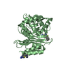
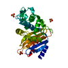
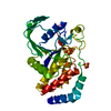
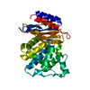
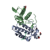
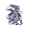
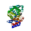
| Title | Crystal structure of the class A beta-lactamse L2 from Stenotrophomonas maltophilia at 1.51 angstrom | ||||||
 Components Components | L2 BETA LACTAMASE | ||||||
 Keywords Keywords | HYDROLASE / BETA-LACTAMASE / CLASS A / L2 | ||||||
| Function / homology |  Function and homology information Function and homology informationbeta-lactam antibiotic catabolic process / beta-lactamase activity / beta-lactamase / response to antibiotic Similarity search - Function | ||||||
| Biological species |  STENOTROPHOMONAS MALTOPHILIA (bacteria) STENOTROPHOMONAS MALTOPHILIA (bacteria) | ||||||
| Method |  X-RAY DIFFRACTION / SYNCHROTRON / MOLECULAR REPLACEMENT / Resolution: 1.51 Å X-RAY DIFFRACTION / SYNCHROTRON / MOLECULAR REPLACEMENT / Resolution: 1.51 Å | ||||||
 Authors Authors | Pernot, L. / Petrella, S. / Sougakoff, W. | ||||||
 Citation Citation | Journal: To be Published Title: Role of the Disulfide Bridge Cys69-Cys238 in Class a B-Lactamases : A Structural and Biochemical Investigation on the B-Lactamase L2 from Stenotrophomonas Maltophilia Authors: Petrella, S. / Pernot, L. / Lascoux, D. / Forest, E. / Jarlier, V. / Sougakoff, W. | ||||||
| History |
|
- Structure visualization
| Structure viewer | Molecule: MolmilJmol/JSmol |
|---|
- Downloads & links
-Download
| PDBx/mmCIF format | 1o7e.cif.gz | 132 KB | Display | PDBx/mmCIF format |
|---|---|---|---|---|
| PDB format | pdb1o7e.ent.gz | 102.9 KB | Display | PDB format |
| PDBx/mmJSON format | 1o7e.json.gz | Tree view | PDBx/mmJSON format | |
| Others |  Other downloads Other downloads |
-Validation report
| Arichive directory | https://data.pdbj.org/pub/pdb/validation_reports/o7/1o7eftp://data.pdbj.org/pub/pdb/validation_reports/o7/1o7e | HTTPS FTP |
|---|
-Related structure data
| Related structure data |  1n4oS S: Starting model for refinement |
|---|---|
| Similar structure data |


-Links
 PDBj
PDBj


- Assembly
| Deposited unit | 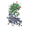
| ||||||||
|---|---|---|---|---|---|---|---|---|---|
| 1 | 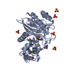
| ||||||||
| 2 | 
| ||||||||
| Unit cell |
| ||||||||
| Noncrystallographic symmetry (NCS) | NCS oper: (Code: given Matrix: (-0.885043, -0.448562, 0.124462), Vector: |
-Components
| #1: Protein | Mass: 29164.059 Da / Num. of mol.: 2 Source method: isolated from a genetically manipulated source Source: (gene. exp.) STENOTROPHOMONAS MALTOPHILIA (bacteria)Strain: 405 / Plasmid: PET29-(A+) / Production host: #2: Chemical |   Mass: 92.094 Da / Num. of mol.: 3 / Source method: obtained synthetically / Formula: C3H8O3 Mass: 92.094 Da / Num. of mol.: 3 / Source method: obtained synthetically / Formula: C3H8O3#3: Chemical | ChemComp-SO4 /   Mass: 96.063 Da / Num. of mol.: 13 / Source method: obtained synthetically / Formula: SO4 Mass: 96.063 Da / Num. of mol.: 13 / Source method: obtained synthetically / Formula: SO4#4: Water | ChemComp-HOH / |  Mass: 18.015 Da / Num. of mol.: 733 / Source method: isolated from a natural source / Formula: H2O Mass: 18.015 Da / Num. of mol.: 733 / Source method: isolated from a natural source / Formula: H2OSequence details | THE L2 MATURE PROTEIN STARTS AT RESIDUE 28 THERE ARE NO RESIDUES NUMBERED 58 AND 239 | |
|---|
-Experimental details
-Experiment
| Experiment | Method: X-RAY DIFFRACTION / Number of used crystals: 1 |
|---|
- Sample preparation
| Crystal | Density Matthews: 3.57 Å3/Da / Density % sol: 65.23 % |
|---|---|
| Crystal grow | pH: 6.5 Details: AMMONIUM SULFATE 1.5M, SODIUM CACODYLATE 0.1M PH 6.5 |
-Data collection
| Diffraction | Mean temperature: 100 K |
|---|---|
| Diffraction source | Source: SYNCHROTRON / Site: LURE  / Beamline: DW32 / Wavelength: 0.948 / Beamline: DW32 / Wavelength: 0.948 |
| Detector | Type: MARRESEARCH / Detector: IMAGE PLATE / Date: Jun 25, 2001 |
| Radiation | Monochromator: SI(111) / Protocol: SINGLE WAVELENGTH / Monochromatic (M) / Laue (L): M / Scattering type: x-ray |
| Radiation wavelength | Wavelength: 0.948 Å / Relative weight: 1 |
| Reflection | Resolution: 1.51→23.9 Å / Num. obs: 131717 / % possible obs: 99.4 % / Observed criterion σ(I): 2.5 / Redundancy: 4.8 % / Biso Wilson estimate: 19.9 Å2 / Rmerge(I) obs: 0.046 / Net I/σ(I): 22.7 |
| Reflection shell | Resolution: 1.51→1.59 Å / Redundancy: 4.6 % / Rmerge(I) obs: 0.17 / Mean I/σ(I) obs: 4.9 / % possible all: 96 |
- Processing
| Software |
| ||||||||||||||||||||
|---|---|---|---|---|---|---|---|---|---|---|---|---|---|---|---|---|---|---|---|---|---|
| Refinement | Method to determine structure: MOLECULAR REPLACEMENT Starting model: THE STARTING MODEL USED WAS THE ONE OF L2 AT 1.85 ANGSTROM, PDB ENTRY 1N4O Resolution: 1.51→23.92 Å / SU B: 0.9034 / SU ML: 0.0346 / Cross valid method: THROUGHOUT / ESU R: 0.14 / ESU R Free: 0.14
| ||||||||||||||||||||
| Displacement parameters | Biso mean: 13.9 Å2 | ||||||||||||||||||||
| Refinement step | Cycle: LAST / Resolution: 1.51→23.92 Å
|
