 Movie
Movie Controller

[English] 日本語
 Yorodumi
- PDB-8ipi: The apo structure of human mitochondrial methyltransferase METTL15 -
+ Open data

- Basic information
| Entry | Database: PDB / ID: 8ipi | ||||||
|---|---|---|---|---|---|---|---|
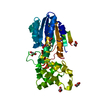
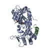
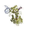
| Title | The apo structure of human mitochondrial methyltransferase METTL15 | ||||||
 Components Components | 12S rRNA N4-methylcytidine (m4C) methyltransferase | ||||||
 Keywords Keywords | RIBOSOMAL PROTEIN / human mitochondrial methyltransferase METTL15 | ||||||
| Function / homology |  Function and homology information Function and homology informationrRNA (cytosine-N4-)-methyltransferase activity / mitochondrial small ribosomal subunit assembly / rRNA base methylation / Transferases; Transferring one-carbon groups; Methyltransferases / mitochondrial matrix / mitochondrion Similarity search - Function | ||||||
| Biological species |  Homo sapiens (human) Homo sapiens (human) | ||||||
| Method |  X-RAY DIFFRACTION / SYNCHROTRON / MOLECULAR REPLACEMENT / Resolution: 2.1 Å X-RAY DIFFRACTION / SYNCHROTRON / MOLECULAR REPLACEMENT / Resolution: 2.1 Å | ||||||
 Authors Authors | Lv, M.Q. / Zhou, W.W. | ||||||
| Funding support |  China, 1items China, 1items
| ||||||
 Citation Citation | Journal: Cell Discov / Year: 2024 Title: Structural insights into the specific recognition of mitochondrial ribosome-binding factor hsRBFA and 12 S rRNA by methyltransferase METTL15. Authors: Lv, M. / Zhou, W. / Hao, Y. / Li, F. / Zhang, H. / Yao, X. / Shi, Y. / Zhang, L. | ||||||
| History |
|
- Structure visualization
| Structure viewer | Molecule: MolmilJmol/JSmol |
|---|
- Downloads & links
-Download
| PDBx/mmCIF format | 8ipi.cif.gz | 129.5 KB | Display | PDBx/mmCIF format |
|---|---|---|---|---|
| PDB format | pdb8ipi.ent.gz | 99.7 KB | Display | PDB format |
| PDBx/mmJSON format | 8ipi.json.gz | Tree view | PDBx/mmJSON format | |
| Others |  Other downloads Other downloads |
-Validation report
| Arichive directory | https://data.pdbj.org/pub/pdb/validation_reports/ip/8ipiftp://data.pdbj.org/pub/pdb/validation_reports/ip/8ipi | HTTPS FTP |
|---|
-Related structure data
-Links
 PDBj
PDBj- Assembly
| Deposited unit | 
| ||||||||
|---|---|---|---|---|---|---|---|---|---|
| 1 |
| ||||||||
| Unit cell |
|
-Components
| #1: Protein | Mass: 37768.621 Da / Num. of mol.: 1 Source method: isolated from a genetically manipulated source Source: (gene. exp.) Homo sapiens (human) / Gene: METTL15, METT5D1 / Production host:  References: UniProt: A6NJ78, Transferases; Transferring one-carbon groups; Methyltransferases |
|---|---|
| #2: Water | ChemComp-HOH /  Mass: 18.015 Da / Num. of mol.: 110 / Source method: isolated from a natural source / Formula: H2O Mass: 18.015 Da / Num. of mol.: 110 / Source method: isolated from a natural source / Formula: H2O |
-Experimental details
-Experiment
| Experiment | Method: X-RAY DIFFRACTION / Number of used crystals: 1 |
|---|
- Sample preparation
| Crystal | Density Matthews: 2.59 Å3/Da / Density % sol: 52.55 % |
|---|---|
| Crystal grow | Temperature: 289 K / Method: vapor diffusion, sitting drop Details: 10% PEG MME 2000, 0.2M Ammonium sulfate, 0.1 M sodium acetate (pH 5.5) |
-Data collection
| Diffraction | Mean temperature: 100 K / Serial crystal experiment: N |
|---|---|
| Diffraction source | Source: SYNCHROTRON / Site: SSRF / Beamline: BL19U1 / Wavelength: 0.979 Å |
| Detector | Type: DECTRIS PILATUS3 6M / Detector: PIXEL / Date: Jul 24, 2022 |
| Radiation | Protocol: SINGLE WAVELENGTH / Monochromatic (M) / Laue (L): M / Scattering type: x-ray |
| Radiation wavelength | Wavelength: 0.979 Å / Relative weight: 1 |
| Reflection | Resolution: 2.1→31.34 Å / Num. obs: 23694 / % possible obs: 100 % / Redundancy: 17.9 % / Rmerge(I) obs: 0.148 / Net I/σ(I): 15.8 |
| Reflection shell | Resolution: 2.1→2.14 Å / Rmerge(I) obs: 0.8 / Mean I/σ(I) obs: 2.8 / Num. unique obs: 1134 |
- Processing
| Software |
| |||||||||||||||||||||||||||||||||||||||||||||||||||||||||||||||
|---|---|---|---|---|---|---|---|---|---|---|---|---|---|---|---|---|---|---|---|---|---|---|---|---|---|---|---|---|---|---|---|---|---|---|---|---|---|---|---|---|---|---|---|---|---|---|---|---|---|---|---|---|---|---|---|---|---|---|---|---|---|---|---|---|
| Refinement | Method to determine structure: MOLECULAR REPLACEMENT / Resolution: 2.1→31.336 Å / SU ML: 0.21 / Cross valid method: FREE R-VALUE / σ(F): 1.35 / Phase error: 21.82 / Stereochemistry target values: ML
| |||||||||||||||||||||||||||||||||||||||||||||||||||||||||||||||
| Solvent computation | Shrinkage radii: 0.9 Å / VDW probe radii: 1.11 Å / Solvent model: FLAT BULK SOLVENT MODEL | |||||||||||||||||||||||||||||||||||||||||||||||||||||||||||||||
| Refinement step | Cycle: LAST / Resolution: 2.1→31.336 Å
| |||||||||||||||||||||||||||||||||||||||||||||||||||||||||||||||
| Refine LS restraints |
| |||||||||||||||||||||||||||||||||||||||||||||||||||||||||||||||
| LS refinement shell |
| |||||||||||||||||||||||||||||||||||||||||||||||||||||||||||||||
| Refinement TLS params. | Method: refined / Origin x: 26.4696 Å / Origin y: -26.3576 Å / Origin z: -7.8066 Å
| |||||||||||||||||||||||||||||||||||||||||||||||||||||||||||||||
| Refinement TLS group | Selection details: (chain 'A' and resid 8 through 338) |
