 Movie
Movie Controller

[English] 日本語
 Yorodumi
- PDB-8ihp: Structure of Semliki Forest virus VLP in complex with the recepto... -
+ Open data

- Basic information
| Entry | Database: PDB / ID: 8ihp | |||||||||||||||||||||||||||||||||||||||||||||||||||||||||||||||||||||
|---|---|---|---|---|---|---|---|---|---|---|---|---|---|---|---|---|---|---|---|---|---|---|---|---|---|---|---|---|---|---|---|---|---|---|---|---|---|---|---|---|---|---|---|---|---|---|---|---|---|---|---|---|---|---|---|---|---|---|---|---|---|---|---|---|---|---|---|---|---|---|
| Title | Structure of Semliki Forest virus VLP in complex with the receptor VLDLR-LA3 | |||||||||||||||||||||||||||||||||||||||||||||||||||||||||||||||||||||
 Components Components |
| |||||||||||||||||||||||||||||||||||||||||||||||||||||||||||||||||||||
 Keywords Keywords | VIRAL PROTEIN / Semliki Forest virus / SFV / receptor / complex / VLDLR / glycoprotein | |||||||||||||||||||||||||||||||||||||||||||||||||||||||||||||||||||||
| Function / homology |  Function and homology information Function and homology informationreelin receptor activity / VLDL clearance / glycoprotein transport / very-low-density lipoprotein particle receptor activity / ventral spinal cord development / Reelin signalling pathway / very-low-density lipoprotein particle binding / low-density lipoprotein particle receptor activity / very-low-density lipoprotein particle clearance / togavirin ...reelin receptor activity / VLDL clearance / glycoprotein transport / very-low-density lipoprotein particle receptor activity / ventral spinal cord development / Reelin signalling pathway / very-low-density lipoprotein particle binding / low-density lipoprotein particle receptor activity / very-low-density lipoprotein particle clearance / togavirin / reelin-mediated signaling pathway / very-low-density lipoprotein particle / positive regulation of dendrite development / cargo receptor activity / T=4 icosahedral viral capsid / lipid transport / dendrite morphogenesis / virion assembly / regulation of synapse assembly / small molecule binding / apolipoprotein binding / cholesterol metabolic process / clathrin-coated pit / receptor-mediated endocytosis / VLDLR internalisation and degradation / memory / calcium-dependent protein binding / nervous system development / host cell endosome / symbiont-mediated suppression of host toll-like receptor signaling pathway / clathrin-dependent endocytosis of virus by host cell / receptor complex / viral translational frameshifting / lysosomal membrane / serine-type endopeptidase activity / fusion of virus membrane with host endosome membrane / viral envelope / calcium ion binding / virion attachment to host cell / host cell nucleus / host cell plasma membrane / glutamatergic synapse / virion membrane / structural molecule activity / signal transduction / proteolysis / RNA binding / membrane / plasma membrane Similarity search - Function | |||||||||||||||||||||||||||||||||||||||||||||||||||||||||||||||||||||
| Biological species |  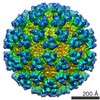 Semliki Forest virus Semliki Forest virus Homo sapiens (human) Homo sapiens (human) | |||||||||||||||||||||||||||||||||||||||||||||||||||||||||||||||||||||
| Method | ELECTRON MICROSCOPY / single particle reconstruction / cryo EM / Resolution: 3 Å | |||||||||||||||||||||||||||||||||||||||||||||||||||||||||||||||||||||
 Authors Authors | Cao, D. / Ma, B. / Cao, Z. / Zhang, X. / Xiang, Y. | |||||||||||||||||||||||||||||||||||||||||||||||||||||||||||||||||||||
| Funding support |  China, 3items China, 3items
| |||||||||||||||||||||||||||||||||||||||||||||||||||||||||||||||||||||
 Citation Citation | Journal: Cell / Year: 2023 Title: Structure of Semliki Forest virus in complex with its receptor VLDLR. Authors: Duanfang Cao / Bingting Ma / Ziyi Cao / Xinzheng Zhang / Ye Xiang / Abstract: Semliki Forest virus (SFV) is an alphavirus that uses the very-low-density lipoprotein receptor (VLDLR) as a receptor during infection of its vertebrate hosts and insect vectors. Herein, we used ...Semliki Forest virus (SFV) is an alphavirus that uses the very-low-density lipoprotein receptor (VLDLR) as a receptor during infection of its vertebrate hosts and insect vectors. Herein, we used cryoelectron microscopy to study the structure of SFV in complex with VLDLR. We found that VLDLR binds multiple E1-DIII sites of SFV through its membrane-distal LDLR class A (LA) repeats. Among the LA repeats of the VLDLR, LA3 has the best binding affinity to SFV. The high-resolution structure shows that LA3 binds SFV E1-DIII through a small surface area of 378 Å, with the main interactions at the interface involving salt bridges. Compared with the binding of single LA3s, consecutive LA repeats around LA3 promote synergistic binding to SFV, during which the LAs undergo a rotation, allowing simultaneous key interactions at multiple E1-DIII sites on the virion and enabling the binding of VLDLRs from divergent host species to SFV. | |||||||||||||||||||||||||||||||||||||||||||||||||||||||||||||||||||||
| History |
|
- Structure visualization
| Structure viewer | Molecule: MolmilJmol/JSmol |
|---|
- Downloads & links
-Download
| PDBx/mmCIF format | 8ihp.cif.gz | 709 KB | Display | PDBx/mmCIF format |
|---|---|---|---|---|
| PDB format | pdb8ihp.ent.gz | Display | PDB format | |
| PDBx/mmJSON format | 8ihp.json.gz | Tree view | PDBx/mmJSON format | |
| Others |  Other downloads Other downloads |
-Validation report
| Summary document | 8ihp_validation.pdf.gz | 464.6 KB | Display | wwPDB validaton report |
|---|---|---|---|---|
| Full document | 8ihp_full_validation.pdf.gz | 506.8 KB | Display | |
| Data in XML | 8ihp_validation.xml.gz | 80.7 KB | Display | |
| Data in CIF | 8ihp_validation.cif.gz | 123.5 KB | Display | |
| Arichive directory | https://data.pdbj.org/pub/pdb/validation_reports/ih/8ihpftp://data.pdbj.org/pub/pdb/validation_reports/ih/8ihp | HTTPS FTP |
-Related structure data
| Related structure data |  35451MC M: map data used to model this data C: citing same article ( |
|---|---|
| Similar structure data |


-Links
 PDBj
PDBj


- Assembly
| Deposited unit | 
|
|---|---|
| 1 |
|
-Components
-Spike glycoprotein ... , 2 types, 8 molecules ADGJBEHK
| #1: Protein | Mass: 46870.277 Da / Num. of mol.: 4 Source method: isolated from a genetically manipulated source Source: (gene. exp.) Semliki Forest virus / Production host: Homo sapiens (human) / References: UniProt: P03315#2: Protein | Mass: 47489.766 Da / Num. of mol.: 4 Source method: isolated from a genetically manipulated source Source: (gene. exp.) Semliki Forest virus / Production host: Homo sapiens (human) / References: UniProt: P03315 |
|---|
-Protein / Protein/peptide / Sugars / Non-polymers , 4 types, 22 molecules CFILMNO

| #3: Protein | Mass: 17848.350 Da / Num. of mol.: 4 Source method: isolated from a genetically manipulated source Source: (gene. exp.) Semliki Forest virus / Production host: Homo sapiens (human) / References: UniProt: P03315, togavirin#4: Protein/peptide | Mass: 4361.682 Da / Num. of mol.: 3 Source method: isolated from a genetically manipulated source Source: (gene. exp.) Homo sapiens (human) / Gene: VLDLR / Production host: Homo sapiens (human) / References: UniProt: P98155#5: Sugar | ChemComp-NAG /  Type: D-saccharide, beta linking / Mass: 221.208 Da / Num. of mol.: 12 / Source method: obtained synthetically / Formula: C8H15NO6 Type: D-saccharide, beta linking / Mass: 221.208 Da / Num. of mol.: 12 / Source method: obtained synthetically / Formula: C8H15NO6#6: Chemical |  Mass: 40.078 Da / Num. of mol.: 3 / Source method: obtained synthetically / Formula: Ca Mass: 40.078 Da / Num. of mol.: 3 / Source method: obtained synthetically / Formula: Ca |
|---|
-Details
| Has ligand of interest | N |
|---|---|
| Has protein modification | Y |
-Experimental details
-Experiment
| Experiment | Method: ELECTRON MICROSCOPY |
|---|---|
| EM experiment | Aggregation state: PARTICLE / 3D reconstruction method: single particle reconstruction |
- Sample preparation
| Component | Name: Semliki Forest virus VLP in complex with its receptor VLDLR at the asymmetric unit Type: COMPLEX / Entity ID: #1-#4 / Source: MULTIPLE SOURCES |
|---|---|
| Source (natural) | Organism: Semliki Forest virus |
| Source (recombinant) | Organism: Homo sapiens (human) |
| Buffer solution | pH: 8 |
| Specimen | Embedding applied: NO / Shadowing applied: NO / Staining applied: NO / Vitrification applied: YES |
| Vitrification | Cryogen name: ETHANE |
- Electron microscopy imaging
| Experimental equipment |  Model: Titan Krios / Image courtesy: FEI Company |
|---|---|
| Microscopy | Model: FEI TITAN KRIOS |
| Electron gun | Electron source:  FIELD EMISSION GUN / Accelerating voltage: 300 kV / Illumination mode: FLOOD BEAM FIELD EMISSION GUN / Accelerating voltage: 300 kV / Illumination mode: FLOOD BEAM |
| Electron lens | Mode: BRIGHT FIELD / Nominal defocus max: 1700 nm / Nominal defocus min: 1200 nm |
| Image recording | Electron dose: 50 e/Å2 / Film or detector model: GATAN K2 SUMMIT (4k x 4k) |
- Processing
| Software | Name: PHENIX / Version: 1.18.2_3874: / Classification: refinement | ||||||||||||||||||||||||
|---|---|---|---|---|---|---|---|---|---|---|---|---|---|---|---|---|---|---|---|---|---|---|---|---|---|
| EM software | Name: PHENIX / Category: model refinement | ||||||||||||||||||||||||
| CTF correction | Type: PHASE FLIPPING AND AMPLITUDE CORRECTION | ||||||||||||||||||||||||
| 3D reconstruction | Resolution: 3 Å / Resolution method: FSC 0.143 CUT-OFF / Num. of particles: 651086 / Symmetry type: POINT | ||||||||||||||||||||||||
| Refine LS restraints |
|
