 Movie
Movie Controller


+ Open data

- Basic information
| Entry | Database: PDB / ID: 8ifb | |||||||||||||||||||||||||||||||||||||||||||||||||||||||||
|---|---|---|---|---|---|---|---|---|---|---|---|---|---|---|---|---|---|---|---|---|---|---|---|---|---|---|---|---|---|---|---|---|---|---|---|---|---|---|---|---|---|---|---|---|---|---|---|---|---|---|---|---|---|---|---|---|---|---|
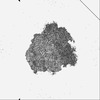
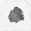
| Title | Dibekacin-bound E.coli 70S ribosome in the PURE system | |||||||||||||||||||||||||||||||||||||||||||||||||||||||||
 Components Components |
| |||||||||||||||||||||||||||||||||||||||||||||||||||||||||
 Keywords Keywords | RIBOSOME | |||||||||||||||||||||||||||||||||||||||||||||||||||||||||
| Function / homology |  Function and homology information Function and homology informationnegative regulation of cytoplasmic translational initiation / transcription antitermination factor activity, RNA binding / ornithine decarboxylase inhibitor activity / misfolded RNA binding / Group I intron splicing / RNA folding / transcriptional attenuation / endoribonuclease inhibitor activity / positive regulation of ribosome biogenesis / RNA-binding transcription regulator activity ...negative regulation of cytoplasmic translational initiation / transcription antitermination factor activity, RNA binding / ornithine decarboxylase inhibitor activity / misfolded RNA binding / Group I intron splicing / RNA folding / transcriptional attenuation / endoribonuclease inhibitor activity / positive regulation of ribosome biogenesis / RNA-binding transcription regulator activity / four-way junction DNA binding / negative regulation of cytoplasmic translation / DnaA-L2 complex / regulation of mRNA stability / translation repressor activity / negative regulation of translational initiation / negative regulation of DNA-templated DNA replication initiation / mRNA regulatory element binding translation repressor activity / positive regulation of RNA splicing / regulation of DNA-templated transcription elongation / transcription elongation factor complex / response to reactive oxygen species / cytosolic ribosome assembly / ribosome assembly / assembly of large subunit precursor of preribosome / transcription antitermination / DNA endonuclease activity / translational initiation / regulation of cell growth / DNA-templated transcription termination / response to radiation / maintenance of translational fidelity / mRNA 5'-UTR binding / regulation of translation / large ribosomal subunit / transferase activity / ribosomal small subunit assembly / ribosome biogenesis / ribosome binding / ribosomal small subunit biogenesis / 5S rRNA binding / ribosomal large subunit assembly / small ribosomal subunit / small ribosomal subunit rRNA binding / large ribosomal subunit rRNA binding / cytosolic small ribosomal subunit / cytosolic large ribosomal subunit / cytoplasmic translation / tRNA binding / negative regulation of translation / rRNA binding / structural constituent of ribosome / ribosome / translation / response to antibiotic / negative regulation of DNA-templated transcription / hydrolase activity / mRNA binding / DNA binding / RNA binding / zinc ion binding / membrane / cytoplasm / cytosol Similarity search - Function | |||||||||||||||||||||||||||||||||||||||||||||||||||||||||
| Biological species |  | |||||||||||||||||||||||||||||||||||||||||||||||||||||||||
| Method | ELECTRON MICROSCOPY / single particle reconstruction / cryo EM / Resolution: 2.43 Å | |||||||||||||||||||||||||||||||||||||||||||||||||||||||||
 Authors Authors | Tomono, J. / Asano, K. / Chiashi, T. / Tanaka, Y. / Yokoyama, T. | |||||||||||||||||||||||||||||||||||||||||||||||||||||||||
| Funding support |  Japan, 2items Japan, 2items
| |||||||||||||||||||||||||||||||||||||||||||||||||||||||||
 Citation Citation | Journal: J Biochem / Year: 2024 Title: Direct visualization of ribosomes in the cell-free system revealed the functional evolution of aminoglycoside. Authors: Junta Tomono / Kosuke Asano / Takuma Chiashi / Masato Suzuki / Masayuki Igarashi / Yoshiaki Takahashi / Yoshikazu Tanaka / Takeshi Yokoyama / Abstract: The rapid emergence of multi-drug-resistant bacteria has raised a serious public health concern. Therefore, new antibiotic developments have been highly desired. Here, we propose a new method to ...The rapid emergence of multi-drug-resistant bacteria has raised a serious public health concern. Therefore, new antibiotic developments have been highly desired. Here, we propose a new method to visualize antibiotic actions on translating ribosomes in the cell-free system under macromolecular crowding conditions by cryo-electron microscopy, designated as the DARC method: the Direct visualization of Antibiotic binding on Ribosomes in the Cell-free translation system. This new method allows for acquiring a more comprehensive understanding of the mode of action of antibiotics on the translation inhibition without ribosome purification. Furthermore, with the direct link to biochemical analysis at the same condition as cryo-EM observation, we revealed the evolution of 2-DOS aminoglycosides from dibekacin (DBK) to arbekacin (ABK) by acquiring the synthetic tailored anchoring motif to lead to stronger binding affinity to ribosomes. Our cryo-EM structures of DBK and ABK bound ribosomes in the cell-free environment clearly depicted a synthetic tailored γ-amino-α-hydroxybutyryl (HABA) motif formed additional interactions with the ribosome enhancing antibiotic bindings. This new approach would be valuable for understanding the function of antibiotics for more efficient drug development. | |||||||||||||||||||||||||||||||||||||||||||||||||||||||||
| History |
|
- Structure visualization
| Structure viewer | Molecule: MolmilJmol/JSmol |
|---|
- Downloads & links
-Download
| PDBx/mmCIF format | 8ifb.cif.gz | 4.3 MB | Display | PDBx/mmCIF format |
|---|---|---|---|---|
| PDB format | pdb8ifb.ent.gz | Display | PDB format | |
| PDBx/mmJSON format | 8ifb.json.gz | Tree view | PDBx/mmJSON format | |
| Others |  Other downloads Other downloads |
-Validation report
| Arichive directory | https://data.pdbj.org/pub/pdb/validation_reports/if/8ifbftp://data.pdbj.org/pub/pdb/validation_reports/if/8ifb | HTTPS FTP |
|---|
-Related structure data
| Related structure data | 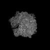 35411MC  8ifcC  8ifdC  8ifeC M: map data used to model this data C: citing same article ( |
|---|---|
| Similar structure data |

-Links
 PDBj
PDBj


- Assembly
| Deposited unit | 
|
|---|---|
| 1 |
|
-Components
-RNA chain , 6 types, 6 molecules Aab567
| #1: RNA chain | Mass: 499873.406 Da / Num. of mol.: 1 / Source method: isolated from a natural source / Source: (natural) |
|---|---|
| #22: RNA chain | Mass: 941811.562 Da / Num. of mol.: 1 / Source method: isolated from a natural source / Source: (natural) |
| #23: RNA chain | Mass: 38790.090 Da / Num. of mol.: 1 / Source method: isolated from a natural source / Source: (natural) |
| #53: RNA chain | Mass: 24644.873 Da / Num. of mol.: 1 / Source method: isolated from a natural source / Source: (natural) |
| #54: RNA chain | Mass: 24832.918 Da / Num. of mol.: 1 / Source method: isolated from a natural source / Source: (natural) |
| #55: RNA chain | Mass: 3161.932 Da / Num. of mol.: 1 / Source method: isolated from a natural source / Source: (natural) |
-30S ribosomal protein ... , 20 types, 20 molecules BCDEFGHIJKLMNOPQRSTU
| #2: Protein | Mass: 26781.670 Da / Num. of mol.: 1 / Source method: isolated from a natural source / Source: (natural) |
|---|---|
| #3: Protein | Mass: 26031.316 Da / Num. of mol.: 1 / Source method: isolated from a natural source / Source: (natural) |
| #4: Protein | Mass: 23514.199 Da / Num. of mol.: 1 / Source method: isolated from a natural source / Source: (natural) |
| #5: Protein | Mass: 17629.398 Da / Num. of mol.: 1 / Source method: isolated from a natural source / Source: (natural) |
| #6: Protein | Mass: 15727.512 Da / Num. of mol.: 1 / Source method: isolated from a natural source / Source: (natural) |
| #7: Protein | Mass: 20055.156 Da / Num. of mol.: 1 / Source method: isolated from a natural source / Source: (natural) |
| #8: Protein | Mass: 14146.557 Da / Num. of mol.: 1 / Source method: isolated from a natural source / Source: (natural) |
| #9: Protein | Mass: 14886.270 Da / Num. of mol.: 1 / Source method: isolated from a natural source / Source: (natural) |
| #10: Protein | Mass: 11755.597 Da / Num. of mol.: 1 / Source method: isolated from a natural source / Source: (natural) |
| #11: Protein | Mass: 13871.959 Da / Num. of mol.: 1 / Source method: isolated from a natural source / Source: (natural) |
| #12: Protein | Mass: 13814.249 Da / Num. of mol.: 1 / Source method: isolated from a natural source / Source: (natural) |
| #13: Protein | Mass: 13128.467 Da / Num. of mol.: 1 / Source method: isolated from a natural source / Source: (natural) |
| #14: Protein | Mass: 11606.560 Da / Num. of mol.: 1 / Source method: isolated from a natural source / Source: (natural) |
| #15: Protein | Mass: 10290.816 Da / Num. of mol.: 1 / Source method: isolated from a natural source / Source: (natural) |
| #16: Protein | Mass: 9207.572 Da / Num. of mol.: 1 / Source method: isolated from a natural source / Source: (natural) |
| #17: Protein | Mass: 9724.491 Da / Num. of mol.: 1 / Source method: isolated from a natural source / Source: (natural) |
| #18: Protein | Mass: 9005.472 Da / Num. of mol.: 1 / Source method: isolated from a natural source / Source: (natural) |
| #19: Protein | Mass: 10455.355 Da / Num. of mol.: 1 / Source method: isolated from a natural source / Source: (natural) |
| #20: Protein | Mass: 9708.464 Da / Num. of mol.: 1 / Source method: isolated from a natural source / Source: (natural) |
| #21: Protein | Mass: 8524.039 Da / Num. of mol.: 1 / Source method: isolated from a natural source / Source: (natural) |
+50S ribosomal protein ... , 29 types, 29 molecules cdefghijklmnopqrstuvwxyz01234
-Non-polymers , 5 types, 436 molecules 


| #56: Chemical | ChemComp-MG /  Mass: 24.305 Da / Num. of mol.: 406 / Source method: obtained synthetically / Formula: Mg Mass: 24.305 Da / Num. of mol.: 406 / Source method: obtained synthetically / Formula: Mg#57: Chemical | ChemComp-SPD /  Mass: 145.246 Da / Num. of mol.: 14 / Source method: obtained synthetically / Formula: C7H19N3 Mass: 145.246 Da / Num. of mol.: 14 / Source method: obtained synthetically / Formula: C7H19N3#58: Chemical | ChemComp-84D /  Mass: 451.515 Da / Num. of mol.: 13 / Source method: obtained synthetically / Formula: C18H37N5O8 / Feature type: SUBJECT OF INVESTIGATION Mass: 451.515 Da / Num. of mol.: 13 / Source method: obtained synthetically / Formula: C18H37N5O8 / Feature type: SUBJECT OF INVESTIGATION#59: Chemical | ChemComp-SPM / |  Mass: 202.340 Da / Num. of mol.: 1 / Source method: obtained synthetically / Formula: C10H26N4 Mass: 202.340 Da / Num. of mol.: 1 / Source method: obtained synthetically / Formula: C10H26N4#60: Chemical |  Mass: 65.409 Da / Num. of mol.: 2 / Source method: obtained synthetically / Formula: Zn Mass: 65.409 Da / Num. of mol.: 2 / Source method: obtained synthetically / Formula: Zn |
|---|
-Details
| Has ligand of interest | Y |
|---|---|
| Has protein modification | Y |
-Experimental details
-Experiment
| Experiment | Method: ELECTRON MICROSCOPY |
|---|---|
| EM experiment | Aggregation state: PARTICLE / 3D reconstruction method: single particle reconstruction |
- Sample preparation
| Component | Name: DBK-bound E.coli 70S ribosome in the PURE system / Type: RIBOSOME / Entity ID: #1-#32, #34-#55 / Source: NATURAL |
|---|---|
| Molecular weight | Value: 2.3 MDa / Experimental value: NO |
| Source (natural) | Organism: |
| Buffer solution | pH: 7.6 |
| Specimen | Embedding applied: NO / Shadowing applied: NO / Staining applied: NO / Vitrification applied: YES |
| Specimen support | Details: covered with a homemade ultra-thin carbon film / Grid material: COPPER / Grid mesh size: 200 divisions/in. / Grid type: Quantifoil R1.2/1.3 |
| Vitrification | Instrument: FEI VITROBOT MARK IV / Cryogen name: ETHANE / Humidity: 100 % / Chamber temperature: 277 K |
- Electron microscopy imaging
| Microscopy | Model: JEOL CRYO ARM 300 |
|---|---|
| Electron gun | Electron source:  FIELD EMISSION GUN / Accelerating voltage: 300 kV / Illumination mode: FLOOD BEAM FIELD EMISSION GUN / Accelerating voltage: 300 kV / Illumination mode: FLOOD BEAM |
| Electron lens | Mode: BRIGHT FIELD / Nominal magnification: 60000 X / Nominal defocus max: 2300 nm / Nominal defocus min: 1100 nm / Cs: 2.7 mm / C2 aperture diameter: 20 µm / Alignment procedure: COMA FREE |
| Specimen holder | Cryogen: NITROGEN / Specimen holder model: JEOL CRYOSPECPORTER |
| Image recording | Electron dose: 40 e/Å2 / Film or detector model: GATAN K3 (6k x 4k) / Num. of real images: 7775 |
| EM imaging optics | Energyfilter name: In-column Omega Filter |
| Image scans | Width: 5760 / Height: 4092 |
- Processing
| Software |
| ||||||||||||||||||||||||
|---|---|---|---|---|---|---|---|---|---|---|---|---|---|---|---|---|---|---|---|---|---|---|---|---|---|
| EM software |
| ||||||||||||||||||||||||
| CTF correction | Type: PHASE FLIPPING AND AMPLITUDE CORRECTION | ||||||||||||||||||||||||
| 3D reconstruction | Resolution: 2.43 Å / Resolution method: FSC 0.143 CUT-OFF / Num. of particles: 180055 / Symmetry type: POINT | ||||||||||||||||||||||||
| Refinement | Cross valid method: NONE Stereochemistry target values: GeoStd + Monomer Library + CDL v1.2 | ||||||||||||||||||||||||
| Displacement parameters | Biso mean: 59.54 Å2 | ||||||||||||||||||||||||
| Refine LS restraints |
|
