 Movie
Movie Controller

[English] 日本語
 Yorodumi
- PDB-8id7: Crystal structure of YbiW in complex with 1,5-anhydroglucitol-6-p... -
+ Open data

- Basic information
| Entry | Database: PDB / ID: 8id7 | ||||||
|---|---|---|---|---|---|---|---|
| Title | Crystal structure of YbiW in complex with 1,5-anhydroglucitol-6-phosphate in Escherichia coli | ||||||
 Components Components | Probable dehydratase YbiW | ||||||
 Keywords Keywords | LYASE / glycyl radical enzyme / YbiW / 1 / 5-anhydroglucitol-6-phosphate | ||||||
| Function / homology |  Function and homology information Function and homology information | ||||||
| Biological species |  | ||||||
| Method |  X-RAY DIFFRACTION / SYNCHROTRON / MOLECULAR REPLACEMENT / Resolution: 2.65 Å X-RAY DIFFRACTION / SYNCHROTRON / MOLECULAR REPLACEMENT / Resolution: 2.65 Å | ||||||
 Authors Authors | Ma, K.L. / Zhang, Y. | ||||||
| Funding support |  China, 1items China, 1items
| ||||||
 Citation Citation | Journal: J.Am.Chem.Soc. / Year: 2024 Title: A Widespread Radical-Mediated Glycolysis Pathway. Authors: Ma, K. / Xue, B. / Chu, R. / Zheng, Y. / Sharma, S. / Jiang, L. / Hu, M. / Xie, Y. / Hu, Y. / Tao, T. / Zhou, Y. / Liu, D. / Li, Z. / Yang, Q. / Chen, Y. / Wu, S. / Tong, Y. / Robinson, R.C. ...Authors: Ma, K. / Xue, B. / Chu, R. / Zheng, Y. / Sharma, S. / Jiang, L. / Hu, M. / Xie, Y. / Hu, Y. / Tao, T. / Zhou, Y. / Liu, D. / Li, Z. / Yang, Q. / Chen, Y. / Wu, S. / Tong, Y. / Robinson, R.C. / Yew, W.S. / Jin, X. / Liu, Y. / Zhao, H. / Ang, E.L. / Wei, Y. / Zhang, Y. | ||||||
| History |
|
- Structure visualization
| Structure viewer | Molecule: MolmilJmol/JSmol |
|---|
- Downloads & links
-Download
| PDBx/mmCIF format | 8id7.cif.gz | 172.5 KB | Display | PDBx/mmCIF format |
|---|---|---|---|---|
| PDB format | pdb8id7.ent.gz | 133.2 KB | Display | PDB format |
| PDBx/mmJSON format | 8id7.json.gz | Tree view | PDBx/mmJSON format | |
| Others |  Other downloads Other downloads |
-Validation report
| Arichive directory | https://data.pdbj.org/pub/pdb/validation_reports/id/8id7ftp://data.pdbj.org/pub/pdb/validation_reports/id/8id7 | HTTPS FTP |
|---|
-Related structure data


-Links
 PDBj
PDBj- Assembly
| Deposited unit | 
| ||||||||
|---|---|---|---|---|---|---|---|---|---|
| 1 | 
| ||||||||
| Unit cell |
|
-Components
| #1: Protein | Mass: 90056.977 Da / Num. of mol.: 1 / Mutation: E114A, E115A, K117A Source method: isolated from a genetically manipulated source Source: (gene. exp.) Gene: ybiW, b0823, JW0807 / Production host: References: UniProt: P75793, Lyases; Carbon-oxygen lyases; Hydro-lyases |
|---|---|
| #2: Sugar | ChemComp-0WK / 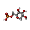  Type: D-saccharide / Mass: 244.136 Da / Num. of mol.: 1 / Source method: obtained synthetically / Formula: C6H13O8P / Feature type: SUBJECT OF INVESTIGATION Type: D-saccharide / Mass: 244.136 Da / Num. of mol.: 1 / Source method: obtained synthetically / Formula: C6H13O8P / Feature type: SUBJECT OF INVESTIGATION |
| #3: Water | ChemComp-HOH /  Mass: 18.015 Da / Num. of mol.: 165 / Source method: isolated from a natural source / Formula: H2O Mass: 18.015 Da / Num. of mol.: 165 / Source method: isolated from a natural source / Formula: H2O |
| Has ligand of interest | Y |
| Has protein modification | N |
-Experimental details
-Experiment
| Experiment | Method: X-RAY DIFFRACTION / Number of used crystals: 1 |
|---|
- Sample preparation
| Crystal | Density Matthews: 3.48 Å3/Da / Density % sol: 64.7 % |
|---|---|
| Crystal grow | Temperature: 291.15 K / Method: vapor diffusion, hanging drop Details: 0.2 M sodium chloride, 0.1M Tris, pH 8.0, 25% (w/v) PEG3350, 10 mM 1,5-anhydroglucitol-6-phosphate |
-Data collection
| Diffraction | Mean temperature: 100 K / Serial crystal experiment: N |
|---|---|
| Diffraction source | Source: SYNCHROTRON / Site: SSRF / Beamline: BL10U2 / Wavelength: 0.9792 Å |
| Detector | Type: DECTRIS EIGER X 16M / Detector: PIXEL / Date: Sep 2, 2022 |
| Radiation | Protocol: SINGLE WAVELENGTH / Monochromatic (M) / Laue (L): M / Scattering type: x-ray |
| Radiation wavelength | Wavelength: 0.9792 Å / Relative weight: 1 |
| Reflection | Resolution: 2.65→57.27 Å / Num. obs: 36549 / % possible obs: 99.9 % / Redundancy: 6.4 % / CC1/2: 0.991 / Rmerge(I) obs: 0.193 / Rpim(I) all: 0.085 / Rrim(I) all: 0.211 / Χ2: 0.99 / Net I/σ(I): 7.7 / Num. measured all: 232578 |
| Reflection shell | Resolution: 2.65→2.72 Å / % possible obs: 99.8 % / Redundancy: 6.8 % / Rmerge(I) obs: 1.589 / Num. measured all: 18068 / Num. unique obs: 2668 / CC1/2: 0.618 / Rpim(I) all: 0.66 / Rrim(I) all: 1.722 / Χ2: 0.82 / Net I/σ(I) obs: 1.3 |
- Processing
| Software |
| |||||||||||||||||||||||||||||||||||||||||||||||||||||||||||||||||||||||||||||||||||||||||||||||||||||||||
|---|---|---|---|---|---|---|---|---|---|---|---|---|---|---|---|---|---|---|---|---|---|---|---|---|---|---|---|---|---|---|---|---|---|---|---|---|---|---|---|---|---|---|---|---|---|---|---|---|---|---|---|---|---|---|---|---|---|---|---|---|---|---|---|---|---|---|---|---|---|---|---|---|---|---|---|---|---|---|---|---|---|---|---|---|---|---|---|---|---|---|---|---|---|---|---|---|---|---|---|---|---|---|---|---|---|---|
| Refinement | Method to determine structure: MOLECULAR REPLACEMENT / Resolution: 2.65→46.482 Å / SU ML: 0.5 / Cross valid method: FREE R-VALUE / σ(F): 0 / Phase error: 32.73 / Stereochemistry target values: ML
| |||||||||||||||||||||||||||||||||||||||||||||||||||||||||||||||||||||||||||||||||||||||||||||||||||||||||
| Solvent computation | Shrinkage radii: 0.9 Å / VDW probe radii: 1.11 Å / Solvent model: FLAT BULK SOLVENT MODEL | |||||||||||||||||||||||||||||||||||||||||||||||||||||||||||||||||||||||||||||||||||||||||||||||||||||||||
| Refinement step | Cycle: LAST / Resolution: 2.65→46.482 Å
| |||||||||||||||||||||||||||||||||||||||||||||||||||||||||||||||||||||||||||||||||||||||||||||||||||||||||
| Refine LS restraints |
| |||||||||||||||||||||||||||||||||||||||||||||||||||||||||||||||||||||||||||||||||||||||||||||||||||||||||
| LS refinement shell |
|
