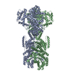
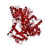
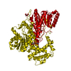
- PDB-8i6z: Crystal structure of apo-form of malonyl-CoA reductase C-domain f... -
+
Open data
ID or keywords:
Loading...
-
Basic information
Entry
Database: PDB / ID: 8i6z
Title
Crystal structure of apo-form of malonyl-CoA reductase C-domain from Chloroflexus aurantiacus
Components
Short-chain dehydrogenase/reductase SDR
Keywords
OXIDOREDUCTASE / NAD(P)-binding protein / Short-chain reductase
Function / homology
Function and homology information
fatty acid elongation / oxidoreductase activity, acting on the CH-OH group of donors, NAD or NADP as acceptor / nucleotide binding / metal ion binding Similarity search - Function
Journal: Int J Biol Macromol / Year: 2023 Title: Cryo-EM structure of bifunctional malonyl-CoA reductase from Chloroflexus aurantiacus reveals a dynamic domain movement for high enzymatic activity. Authors: Jae-Woo Ahn / Sangwoo Kim / Jiyeon Hong / Kyung-Jin Kim / Abstract: The platform chemical 3-hydroxypropionic acid is used to synthesize various valuable materials, including bioplastics. Bifunctional malonyl-CoA reductase is a key enzyme in 3-hydroxypropionic acid ...The platform chemical 3-hydroxypropionic acid is used to synthesize various valuable materials, including bioplastics. Bifunctional malonyl-CoA reductase is a key enzyme in 3-hydroxypropionic acid biosynthesis as it catalyzes the two-step reduction of malonyl-CoA to malonate semialdehyde to 3-hydroxypropionic acid. Here, we report the cryo-EM structure of a full-length malonyl-CoA reductase protein from Chloroflexus aurantiacus (CaMCR). The EM model of CaMCR reveals a tandem helix architecture comprising an N-terminal (CaMCR) and a C-terminal (CaMCR) domain. The CaMCR model also revealed that the enzyme undergoes a dynamic domain movement between CaMCR and CaMCR due to the presence of a flexible linker between these two domains. Increasing the flexibility and extension of the linker resulted in a twofold increase in enzyme activity, indicating that for CaMCR, domain movement is crucial for high enzyme activity. We also describe the structural features of CaMCR and CaMCR. This study reveals the protein structures underlying the molecular mechanism of CaMCR and thereby provides valuable information for future enzyme engineering to improve the productivity of 3-hydroxypropionic acid.
History
Deposition
Jan 30, 2023
Deposition site: PDBJ / Processing site: PDBJ
Revision 1.0
Jun 14, 2023
Provider: repository / Type: Initial release
Revision 1.1
May 29, 2024
Group: Data collection / Category: chem_comp_atom / chem_comp_bond
Method to determine structure: SAD / Resolution: 1.95→26.67 Å / Cor.coef. Fo:Fc: 0.969 / Cor.coef. Fo:Fc free: 0.955 / SU B: 2.723 / SU ML: 0.077 / Cross valid method: THROUGHOUT / ESU R: 0.119 / ESU R Free: 0.114 / Stereochemistry target values: MAXIMUM LIKELIHOOD / Details: HYDROGENS HAVE BEEN ADDED IN THE RIDING POSITIONS
Rfactor
Num. reflection
% reflection
Selection details
Rfree
0.1881
3234
5.2 %
RANDOM
Rwork
0.15643
-
-
-
obs
0.15806
59137
99.37 %
-
Solvent computation
Ion probe radii: 0.8 Å / Shrinkage radii: 0.8 Å / VDW probe radii: 1.2 Å / Solvent model: MASK
 Movie
Movie Controller

 Yorodumi
 Open data
 Basic information
 Components
 Keywords
 Function and homology information
 Chloroflexus aurantiacus (bacteria)
 X-RAY DIFFRACTION /
  Authors
 Citation
 Structure visualization
 Downloads & links
 Other downloads
 PDBj
PDBj


 Assembly
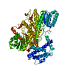

 Mass: 92.094 Da / Num. of mol.: 1 / Source method: obtained synthetically / Formula: C3H8O3
 Mass: 150.087 Da / Num. of mol.: 2 / Source method: obtained synthetically / Formula: C4H6O6 / Feature type: SUBJECT OF INVESTIGATION
 Mass: 18.015 Da / Num. of mol.: 453 / Source method: isolated from a natural source / Formula: H2O
 Sample preparation
 Processing