 Movie
Movie Controller

[English] 日本語
 Yorodumi
- PDB-8htd: Crystal structure of an effector from Chromobacterium violaceum i... -
+ Open data

- Basic information
| Entry | Database: PDB / ID: 8htd | |||||||||||||||||||||
|---|---|---|---|---|---|---|---|---|---|---|---|---|---|---|---|---|---|---|---|---|---|---|
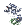
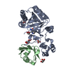
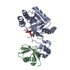
| Title | Crystal structure of an effector from Chromobacterium violaceum in complex with ubiquitin | |||||||||||||||||||||
 Components Components |
| |||||||||||||||||||||
 Keywords Keywords | TRANSFERASE / ubiquitination | |||||||||||||||||||||
| Function / homology |  Function and homology information Function and homology informationglycosyltransferase activity / Transferases; Glycosyltransferases; Pentosyltransferases / ribosomal large subunit export from nucleus / modification-dependent protein catabolic process / protein tag activity / host cell / ribosome biogenesis / ribosomal large subunit assembly / cytosolic large ribosomal subunit / cytoplasmic translation ...glycosyltransferase activity / Transferases; Glycosyltransferases; Pentosyltransferases / ribosomal large subunit export from nucleus / modification-dependent protein catabolic process / protein tag activity / host cell / ribosome biogenesis / ribosomal large subunit assembly / cytosolic large ribosomal subunit / cytoplasmic translation / structural constituent of ribosome / protein ubiquitination / ubiquitin protein ligase binding / extracellular region / nucleus / cytoplasm / cytosol Similarity search - Function | |||||||||||||||||||||
| Biological species |  Chromobacterium violaceum ATCC 12472 (bacteria) Chromobacterium violaceum ATCC 12472 (bacteria) | |||||||||||||||||||||
| Method |  X-RAY DIFFRACTION / SYNCHROTRON / MOLECULAR REPLACEMENT / Resolution: 1.848 Å X-RAY DIFFRACTION / SYNCHROTRON / MOLECULAR REPLACEMENT / Resolution: 1.848 Å | |||||||||||||||||||||
 Authors Authors | Tan, J. / Wang, X. / Zhou, Y. / Zhu, Y. | |||||||||||||||||||||
| Funding support |  China, 6items China, 6items
| |||||||||||||||||||||
 Citation Citation | Journal: Nat.Chem.Biol. / Year: 2024 Title: Molecular basis of threonine ADP-ribosylation of ubiquitin by bacterial ARTs. Authors: Tan, J. / Xu, Y. / Wang, X. / Yan, F. / Xian, W. / Liu, X. / Chen, Y. / Zhu, Y. / Zhou, Y. | |||||||||||||||||||||
| History |
|
- Structure visualization
| Structure viewer | Molecule: MolmilJmol/JSmol |
|---|
- Downloads & links
-Download
| PDBx/mmCIF format | 8htd.cif.gz | 135.4 KB | Display | PDBx/mmCIF format |
|---|---|---|---|---|
| PDB format | pdb8htd.ent.gz | 102.4 KB | Display | PDB format |
| PDBx/mmJSON format | 8htd.json.gz | Tree view | PDBx/mmJSON format | |
| Others |  Other downloads Other downloads |
-Validation report
| Arichive directory | https://data.pdbj.org/pub/pdb/validation_reports/ht/8htdftp://data.pdbj.org/pub/pdb/validation_reports/ht/8htd | HTTPS FTP |
|---|
-Related structure data
-Links
 PDBj
PDBj

- Assembly
| Deposited unit | 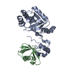
| ||||||||
|---|---|---|---|---|---|---|---|---|---|
| 1 |
| ||||||||
| Unit cell |
|
-Components
| #1: Protein | Mass: 26078.938 Da / Num. of mol.: 1 Source method: isolated from a genetically manipulated source Source: (gene. exp.) Chromobacterium violaceum ATCC 12472 (bacteria)Strain: ATCC 12472 / DSM 30191 / JCM 1249 / NBRC 12614 / NCIMB 9131 / NCTC 9757 Gene: cteC, CV_1467 / Production host: References: UniProt: Q7NY09, Transferases; Glycosyltransferases; Pentosyltransferases |
|---|---|
| #2: Protein | Mass: 8568.769 Da / Num. of mol.: 1 Source method: isolated from a genetically manipulated source Source: (gene. exp.) |
| #3: Water | ChemComp-HOH /  Mass: 18.015 Da / Num. of mol.: 274 / Source method: isolated from a natural source / Formula: H2O Mass: 18.015 Da / Num. of mol.: 274 / Source method: isolated from a natural source / Formula: H2O |
| Has protein modification | Y |
-Experimental details
-Experiment
| Experiment | Method: X-RAY DIFFRACTION / Number of used crystals: 1 |
|---|
- Sample preparation
| Crystal | Density Matthews: 2.06 Å3/Da / Density % sol: 40.36 % |
|---|---|
| Crystal grow | Temperature: 289 K / Method: vapor diffusion, hanging drop / pH: 6.5 Details: 100 mM sodium cacodylate, pH 6.5, 200 mM sodium acetate, 28% to 30% PEG 8000 |
-Data collection
| Diffraction | Mean temperature: 100 K / Serial crystal experiment: N | |||||||||||||||||||||||||||||||||||||||||||||||||||||||||||||||||||||||||||||||||||||||||||||||||||||||||||||||||||||||||||||||||||||||||||||||||||||||||||||||||||||||||||||||||||||||||||||
|---|---|---|---|---|---|---|---|---|---|---|---|---|---|---|---|---|---|---|---|---|---|---|---|---|---|---|---|---|---|---|---|---|---|---|---|---|---|---|---|---|---|---|---|---|---|---|---|---|---|---|---|---|---|---|---|---|---|---|---|---|---|---|---|---|---|---|---|---|---|---|---|---|---|---|---|---|---|---|---|---|---|---|---|---|---|---|---|---|---|---|---|---|---|---|---|---|---|---|---|---|---|---|---|---|---|---|---|---|---|---|---|---|---|---|---|---|---|---|---|---|---|---|---|---|---|---|---|---|---|---|---|---|---|---|---|---|---|---|---|---|---|---|---|---|---|---|---|---|---|---|---|---|---|---|---|---|---|---|---|---|---|---|---|---|---|---|---|---|---|---|---|---|---|---|---|---|---|---|---|---|---|---|---|---|---|---|---|---|---|---|
| Diffraction source | Source: SYNCHROTRON / Site: SSRF / Beamline: BL19U1 / Wavelength: 0.9789 Å | |||||||||||||||||||||||||||||||||||||||||||||||||||||||||||||||||||||||||||||||||||||||||||||||||||||||||||||||||||||||||||||||||||||||||||||||||||||||||||||||||||||||||||||||||||||||||||||
| Detector | Type: DECTRIS PILATUS 6M / Detector: PIXEL / Date: Apr 8, 2018 | |||||||||||||||||||||||||||||||||||||||||||||||||||||||||||||||||||||||||||||||||||||||||||||||||||||||||||||||||||||||||||||||||||||||||||||||||||||||||||||||||||||||||||||||||||||||||||||
| Radiation | Protocol: SINGLE WAVELENGTH / Monochromatic (M) / Laue (L): M / Scattering type: x-ray | |||||||||||||||||||||||||||||||||||||||||||||||||||||||||||||||||||||||||||||||||||||||||||||||||||||||||||||||||||||||||||||||||||||||||||||||||||||||||||||||||||||||||||||||||||||||||||||
| Radiation wavelength | Wavelength: 0.9789 Å / Relative weight: 1 | |||||||||||||||||||||||||||||||||||||||||||||||||||||||||||||||||||||||||||||||||||||||||||||||||||||||||||||||||||||||||||||||||||||||||||||||||||||||||||||||||||||||||||||||||||||||||||||
| Reflection | Resolution: 1.848→50 Å / Num. obs: 22527 / % possible obs: 93.3 % / Redundancy: 3.7 % / Rmerge(I) obs: 0.083 / Rpim(I) all: 0.05 / Rrim(I) all: 0.098 / Χ2: 1.742 / Net I/σ(I): 9.1 | |||||||||||||||||||||||||||||||||||||||||||||||||||||||||||||||||||||||||||||||||||||||||||||||||||||||||||||||||||||||||||||||||||||||||||||||||||||||||||||||||||||||||||||||||||||||||||||
| Reflection shell | Diffraction-ID: 1
|
- Processing
| Software |
| |||||||||||||||||||||||||||||||||||||||||||||||||||||||||||||||||||||||||||
|---|---|---|---|---|---|---|---|---|---|---|---|---|---|---|---|---|---|---|---|---|---|---|---|---|---|---|---|---|---|---|---|---|---|---|---|---|---|---|---|---|---|---|---|---|---|---|---|---|---|---|---|---|---|---|---|---|---|---|---|---|---|---|---|---|---|---|---|---|---|---|---|---|---|---|---|---|
| Refinement | Method to determine structure: MOLECULAR REPLACEMENT / Resolution: 1.848→41.373 Å / SU ML: 0.24 / Cross valid method: THROUGHOUT / σ(F): 1.39 / Phase error: 25.86
| |||||||||||||||||||||||||||||||||||||||||||||||||||||||||||||||||||||||||||
| Solvent computation | Shrinkage radii: 0.9 Å / VDW probe radii: 1.11 Å | |||||||||||||||||||||||||||||||||||||||||||||||||||||||||||||||||||||||||||
| Displacement parameters | Biso max: 79.49 Å2 / Biso mean: 29.1709 Å2 / Biso min: 7.16 Å2 | |||||||||||||||||||||||||||||||||||||||||||||||||||||||||||||||||||||||||||
| Refinement step | Cycle: final / Resolution: 1.848→41.373 Å
| |||||||||||||||||||||||||||||||||||||||||||||||||||||||||||||||||||||||||||
| LS refinement shell | Refine-ID: X-RAY DIFFRACTION / Rfactor Rfree error: 0
| |||||||||||||||||||||||||||||||||||||||||||||||||||||||||||||||||||||||||||
| Refinement TLS params. | Method: refined / Refine-ID: X-RAY DIFFRACTION
| |||||||||||||||||||||||||||||||||||||||||||||||||||||||||||||||||||||||||||
| Refinement TLS group |
|
