 Movie
Movie Controller

[English] 日本語
 Yorodumi
- PDB-8hjt: Crystal Structure of Intracellular B30.2 Domain of VpBTN3 and VpB... -
+ Open data

- Basic information
| Entry | Database: PDB / ID: 8hjt | ||||||
|---|---|---|---|---|---|---|---|
| Title | Crystal Structure of Intracellular B30.2 Domain of VpBTN3 and VpBTN2 in Complex with HMBPP | ||||||
 Components Components |
| ||||||
 Keywords Keywords | IMMUNE SYSTEM / Butyrophilin / Signaling Protein | ||||||
| Function / homology |  Function and homology information Function and homology informationregulation of cytokine production / T cell receptor signaling pathway / signaling receptor binding / external side of plasma membrane Similarity search - Function | ||||||
| Biological species |  | ||||||
| Method |  X-RAY DIFFRACTION / MOLECULAR REPLACEMENT / Resolution: 2.91 Å X-RAY DIFFRACTION / MOLECULAR REPLACEMENT / Resolution: 2.91 Å | ||||||
 Authors Authors | Yang, Y.Y. / Shen, P.P. / Li, X. / Yi, S.M. / Zhang, M.T. / Huang, J.-W. / Chen, C.-C. / Guo, R.-T. | ||||||
| Funding support |  China, 1items China, 1items
| ||||||
 Citation Citation | Journal: Nature / Year: 2023 Title: Phosphoantigens glue butyrophilin 3A1 and 2A1 to activate V gamma 9V delta 2 T cells. Authors: Yuan, L. / Ma, X. / Yang, Y. / Qu, Y. / Li, X. / Zhu, X. / Ma, W. / Duan, J. / Xue, J. / Yang, H. / Huang, J.W. / Yi, S. / Zhang, M. / Cai, N. / Zhang, L. / Ding, Q. / Lai, K. / Liu, C. / ...Authors: Yuan, L. / Ma, X. / Yang, Y. / Qu, Y. / Li, X. / Zhu, X. / Ma, W. / Duan, J. / Xue, J. / Yang, H. / Huang, J.W. / Yi, S. / Zhang, M. / Cai, N. / Zhang, L. / Ding, Q. / Lai, K. / Liu, C. / Zhang, L. / Liu, X. / Yao, Y. / Zhou, S. / Li, X. / Shen, P. / Chang, Q. / Malwal, S.R. / He, Y. / Li, W. / Chen, C. / Chen, C.C. / Oldfield, E. / Guo, R.T. / Zhang, Y. | ||||||
| History |
|
- Structure visualization
| Structure viewer | Molecule: MolmilJmol/JSmol |
|---|
- Downloads & links
-Download
| PDBx/mmCIF format | 8hjt.cif.gz | 202.5 KB | Display | PDBx/mmCIF format |
|---|---|---|---|---|
| PDB format | pdb8hjt.ent.gz | 129.6 KB | Display | PDB format |
| PDBx/mmJSON format | 8hjt.json.gz | Tree view | PDBx/mmJSON format | |
| Others |  Other downloads Other downloads |
-Validation report
| Arichive directory | https://data.pdbj.org/pub/pdb/validation_reports/hj/8hjtftp://data.pdbj.org/pub/pdb/validation_reports/hj/8hjt | HTTPS FTP |
|---|
-Related structure data
| Related structure data |  8igtC 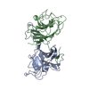 8ih4C 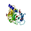 8ixvC 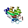 8izeC 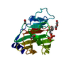 8izgC  8jy9C  8jyaC 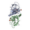 8jybC  8jycC  8jyeC 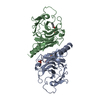 8jyfC C: citing same article ( |
|---|---|
| Similar structure data |
-Links
 PDBj
PDBj
- Assembly
| Deposited unit | 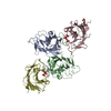
| ||||||||||||
|---|---|---|---|---|---|---|---|---|---|---|---|---|---|
| 1 |
| ||||||||||||
| Unit cell |
|
-Components
| #1: Protein | Mass: 26422.887 Da / Num. of mol.: 2 Source method: isolated from a genetically manipulated source Source: (gene. exp.)  #2: Protein | Mass: 22482.258 Da / Num. of mol.: 2 Source method: isolated from a genetically manipulated source Source: (gene. exp.) #3: Chemical |   Mass: 262.092 Da / Num. of mol.: 2 / Source method: obtained synthetically / Formula: C5H12O8P2 / Feature type: SUBJECT OF INVESTIGATION Mass: 262.092 Da / Num. of mol.: 2 / Source method: obtained synthetically / Formula: C5H12O8P2 / Feature type: SUBJECT OF INVESTIGATION#4: Chemical | ChemComp-EPE / |   Mass: 238.305 Da / Num. of mol.: 1 / Source method: obtained synthetically / Formula: C8H18N2O4S / Comment: pH buffer*YM Mass: 238.305 Da / Num. of mol.: 1 / Source method: obtained synthetically / Formula: C8H18N2O4S / Comment: pH buffer*YM#5: Water | ChemComp-HOH / |  Mass: 18.015 Da / Num. of mol.: 112 / Source method: isolated from a natural source / Formula: H2O Mass: 18.015 Da / Num. of mol.: 112 / Source method: isolated from a natural source / Formula: H2OHas ligand of interest | Y | Has protein modification | N | |
|---|
-Experimental details
-Experiment
| Experiment | Method: X-RAY DIFFRACTION / Number of used crystals: 1 |
|---|
- Sample preparation
| Crystal | Density Matthews: 2.7 Å3/Da / Density % sol: 54.53 % |
|---|---|
| Crystal grow | Temperature: 289 K / Method: vapor diffusion, sitting drop / Details: 12% PEG 8k, 0.1 M HEPES pH7.5 |
-Data collection
| Diffraction | Mean temperature: 100 K / Serial crystal experiment: N |
|---|---|
| Diffraction source | Source: LIQUID ANODE / Type: BRUKER METALJET / Wavelength: 1.34138 Å |
| Detector | Type: Bruker PHOTON III / Detector: PIXEL / Date: Sep 27, 2022 |
| Radiation | Protocol: SINGLE WAVELENGTH / Monochromatic (M) / Laue (L): M / Scattering type: x-ray |
| Radiation wavelength | Wavelength: 1.34138 Å / Relative weight: 1 |
| Reflection | Resolution: 2.91→35 Å / Num. obs: 24182 / % possible obs: 99.1 % / Redundancy: 7.1 % / Biso Wilson estimate: 37.11 Å2 / CC1/2: 1 / Net I/σ(I): 16.9 |
| Reflection shell | Resolution: 2.91→2.96 Å / Num. unique obs: 1131 / CC1/2: 0.96 |
- Processing
| Software |
| |||||||||||||||||||||||||||||||||||||||||||||||||||||||||||||||
|---|---|---|---|---|---|---|---|---|---|---|---|---|---|---|---|---|---|---|---|---|---|---|---|---|---|---|---|---|---|---|---|---|---|---|---|---|---|---|---|---|---|---|---|---|---|---|---|---|---|---|---|---|---|---|---|---|---|---|---|---|---|---|---|---|
| Refinement | Method to determine structure: MOLECULAR REPLACEMENT / Resolution: 2.91→34.66 Å / SU ML: 0.4087 / Cross valid method: FREE R-VALUE / σ(F): 1.38 / Phase error: 25.9585 Stereochemistry target values: GeoStd + Monomer Library + CDL v1.2
| |||||||||||||||||||||||||||||||||||||||||||||||||||||||||||||||
| Solvent computation | Shrinkage radii: 0.9 Å / VDW probe radii: 1.11 Å / Solvent model: FLAT BULK SOLVENT MODEL | |||||||||||||||||||||||||||||||||||||||||||||||||||||||||||||||
| Displacement parameters | Biso mean: 36.64 Å2 | |||||||||||||||||||||||||||||||||||||||||||||||||||||||||||||||
| Refinement step | Cycle: LAST / Resolution: 2.91→34.66 Å
| |||||||||||||||||||||||||||||||||||||||||||||||||||||||||||||||
| Refine LS restraints |
| |||||||||||||||||||||||||||||||||||||||||||||||||||||||||||||||
| LS refinement shell |
|
