Entry Database : PDB / ID : 8hhqTitle Covalent bond formation between cysteine of PPARg-LBD and iodoacetic acid Peroxisome proliferator-activated receptor gamma Keywords / / / / Function / homology Function Domain/homology Component
/ / / / / / / / / / / / / / / / / / / / / / / / / / / / / / / / / / / / / / / / / / / / / / / / / / / / / / / / / / / / / / / / / / / / / / / / / / / / / / / / / / / / / / / / / / / / / / / / / / / / / / / / / / / / / / / / / / / / / / / / / / / / / / / / / / / / / Biological species Homo sapiens (human)Method / / Resolution : 2.4 Å Authors Kojima, H. / Itoh, T. Funding support Organization Grant number Country Japan Agency for Medical Research and Development (AMED)
Journal : J.Med.Chem. / Year : 2023Title : Covalent Modifier Discovery Using Hydrogen/Deuterium Exchange-Mass Spectrometry.Authors : Kojima, H. / Yanagi, R. / Higuchi, E. / Yoshizawa, M. / Shimodaira, T. / Kumagai, M. / Kyoya, T. / Sekine, M. / Egawa, D. / Ohashi, N. / Ishida, H. / Yamamoto, K. / Itoh, T. History Deposition Nov 16, 2022 Deposition site / Processing site Revision 1.0 Sep 20, 2023 Provider / Type Revision 2.0 Nov 15, 2023 Group / Data collection / Derived calculationsCategory atom_site / chem_comp_atom ... atom_site / chem_comp_atom / chem_comp_bond / struct_conn Item _atom_site.auth_atom_id / _atom_site.label_atom_id ... _atom_site.auth_atom_id / _atom_site.label_atom_id / _chem_comp_atom.atom_id / _chem_comp_bond.atom_id_1 / _chem_comp_bond.atom_id_2 / _struct_conn.ptnr2_label_atom_id Revision 2.1 Sep 17, 2025 Group / Derived calculations / Structure summaryCategory pdbx_entry_details / pdbx_unobs_or_zero_occ_atoms ... pdbx_entry_details / pdbx_unobs_or_zero_occ_atoms / pdbx_validate_close_contact / struct_conn Item Revision 2.2 Mar 4, 2026 Group / Category
Show all Show less
 Movie
Movie Controller

 Yorodumi
 Open data
 Basic information
 Components
 Keywords
 Function and homology information
 Homo sapiens (human)
 X-RAY DIFFRACTION /
  Authors
 Japan, 1items
  Citation
 Structure visualization
 Downloads & links
 Other downloads
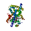
 PDBj
PDBj Assembly
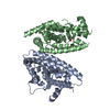

 Mass: 76.051 Da / Num. of mol.: 2 / Source method: obtained synthetically / Formula: C2H4O3 / Feature type: SUBJECT OF INVESTIGATION
 Mass: 18.015 Da / Num. of mol.: 126 / Source method: isolated from a natural source / Formula: H2O
 Sample preparation
 Processing