Entry Database : PDB / ID : 8hhpTitle Crystal structure of PPARg-LBD complexed with three partial agonists, one nTZDpa and two LT175 Peroxisome proliferator-activated receptor gamma Keywords / / / / Function / homology Function Domain/homology Component
/ / / / / / / / / / / / / / / / / / / / / / / / / / / / / / / / / / / / / / / / / / / / / / / / / / / / / / / / / / / / / / / / / / / / / / / / / / / / / / / / / / / / / / / / / / / / / / / / / / / / / / / / / / / / / / / / / / / / / / / / / / / / / / / / / / / / / Biological species Homo sapiens (human)Method / / / Resolution : 2.45 Å Authors Kojima, H. / Itoh, T. Funding support Organization Grant number Country Japan Agency for Medical Research and Development (AMED)
Journal : J.Med.Chem. / Year : 2023Title : Covalent Modifier Discovery Using Hydrogen/Deuterium Exchange-Mass Spectrometry.Authors : Kojima, H. / Yanagi, R. / Higuchi, E. / Yoshizawa, M. / Shimodaira, T. / Kumagai, M. / Kyoya, T. / Sekine, M. / Egawa, D. / Ohashi, N. / Ishida, H. / Yamamoto, K. / Itoh, T. History Deposition Nov 16, 2022 Deposition site / Processing site Revision 1.0 Nov 22, 2023 Provider / Type Revision 1.1 May 22, 2024 Group / Category / citation_authorItem _citation.country / _citation.journal_abbrev ... _citation.country / _citation.journal_abbrev / _citation.journal_id_ASTM / _citation.journal_id_CSD / _citation.journal_id_ISSN / _citation.journal_volume / _citation.page_first / _citation.page_last / _citation.pdbx_database_id_DOI / _citation.pdbx_database_id_PubMed / _citation.title / _citation.year Revision 1.2 Mar 4, 2026 Group / Structure summaryCategory / pdbx_initial_refinement_modelItem
Show all Show less
 Movie
Movie Controller

 Yorodumi
 Open data
 Basic information
 Components
 Keywords
 Function and homology information
 Homo sapiens (human)
 X-RAY DIFFRACTION /
  Authors
 Japan, 1items
  Citation
 Structure visualization
 Downloads & links
 Other downloads
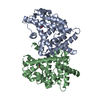
 PDBj
PDBj Assembly

 Mass: 428.331 Da / Num. of mol.: 1 / Source method: obtained synthetically / Formula: C22H15Cl2NO2S
 Mass: 318.366 Da / Num. of mol.: 2 / Source method: obtained synthetically / Formula: C21H18O3 / Feature type: SUBJECT OF INVESTIGATION
 Mass: 18.015 Da / Num. of mol.: 33 / Source method: isolated from a natural source / Formula: H2O
 Sample preparation
 Processing