 Movie
Movie Controller


+ Open data

- Basic information
| Entry | Database: PDB / ID: 8hba | |||||||||
|---|---|---|---|---|---|---|---|---|---|---|
| Title | Crystal structure of NAD-II riboswitch (single strand) with NAD | |||||||||
 Components Components | RNA (55-MER) | |||||||||
 Keywords Keywords | RNA / Riboswitch / NAD / NMN / Aptamer | |||||||||
| Function / homology | NICOTINAMIDE-ADENINE-DINUCLEOTIDE / BETA-NICOTINAMIDE RIBOSE MONOPHOSPHATE / RNA / RNA (> 10) Function and homology information Function and homology information | |||||||||
| Biological species |  Streptococcus parasanguinis (bacteria) Streptococcus parasanguinis (bacteria) | |||||||||
| Method |  X-RAY DIFFRACTION / SYNCHROTRON / MOLECULAR REPLACEMENT / Resolution: 2.64 Å X-RAY DIFFRACTION / SYNCHROTRON / MOLECULAR REPLACEMENT / Resolution: 2.64 Å | |||||||||
 Authors Authors | Peng, X. / Lilley, D.M.J. / Huang, L. | |||||||||
| Funding support |  China, China,  United Kingdom, 2items United Kingdom, 2items
| |||||||||
 Citation Citation | Journal: Nucleic Acids Res. / Year: 2023 Title: Crystal structures of the NAD+-II riboswitch reveal two distinct ligand-binding pockets. Authors: Peng, X. / Liao, W. / Lin, X. / Lilley, D.M.J. / Huang, L. | |||||||||
| History |
|
- Structure visualization
| Structure viewer | Molecule: MolmilJmol/JSmol |
|---|
- Downloads & links
-Download
| PDBx/mmCIF format | 8hba.cif.gz | 124.6 KB | Display | PDBx/mmCIF format |
|---|---|---|---|---|
| PDB format | pdb8hba.ent.gz | 98.6 KB | Display | PDB format |
| PDBx/mmJSON format | 8hba.json.gz | Tree view | PDBx/mmJSON format | |
| Others |  Other downloads Other downloads |
-Validation report
| Arichive directory | https://data.pdbj.org/pub/pdb/validation_reports/hb/8hbaftp://data.pdbj.org/pub/pdb/validation_reports/hb/8hba | HTTPS FTP |
|---|
-Related structure data
| Related structure data |  8hb1SC 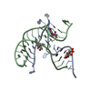 8hb3C 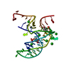 8hb8C 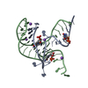 8i3zC S: Starting model for refinement C: citing same article ( |
|---|---|
| Similar structure data |
-Links
 PDBj
PDBj


- Assembly
| Deposited unit | 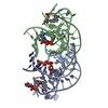
| ||||||||
|---|---|---|---|---|---|---|---|---|---|
| 1 |
| ||||||||
| Unit cell |
|
-Components
| #1: RNA chain | Mass: 18132.947 Da / Num. of mol.: 2 / Source method: obtained synthetically / Source: (synth.) Streptococcus parasanguinis (bacteria)#2: Chemical |   Mass: 663.425 Da / Num. of mol.: 3 / Source method: obtained synthetically / Formula: C21H27N7O14P2 / Feature type: SUBJECT OF INVESTIGATION / Comment: NAD*YM Mass: 663.425 Da / Num. of mol.: 3 / Source method: obtained synthetically / Formula: C21H27N7O14P2 / Feature type: SUBJECT OF INVESTIGATION / Comment: NAD*YM#3: Chemical | ChemComp-NMN / |   Mass: 335.227 Da / Num. of mol.: 1 / Source method: obtained synthetically / Formula: C11H16N2O8P / Feature type: SUBJECT OF INVESTIGATION Mass: 335.227 Da / Num. of mol.: 1 / Source method: obtained synthetically / Formula: C11H16N2O8P / Feature type: SUBJECT OF INVESTIGATIONHas ligand of interest | Y | |
|---|
-Experimental details
-Experiment
| Experiment | Method: X-RAY DIFFRACTION / Number of used crystals: 1 |
|---|
- Sample preparation
| Crystal | Density Matthews: 2.16 Å3/Da / Density % sol: 43.16 % |
|---|---|
| Crystal grow | Temperature: 291 K / Method: vapor diffusion, hanging drop Details: 0.012 M Sodium chloride, 0.08 M Potassium chloride 0.04 M Sodium cacodylate trihydrate pH 5.5 45% v/v (+/-)-2-Methyl-2,4-pentanediol 0.02 M Hexammine cobalt(III) chloride |
-Data collection
| Diffraction | Mean temperature: 100 K / Serial crystal experiment: N |
|---|---|
| Diffraction source | Source: SYNCHROTRON / Site: SSRF / Beamline: BL19U1 / Wavelength: 0.9785 Å |
| Detector | Type: DECTRIS EIGER X 16M / Detector: PIXEL / Date: Sep 20, 2021 |
| Radiation | Protocol: SINGLE WAVELENGTH / Monochromatic (M) / Laue (L): M / Scattering type: x-ray |
| Radiation wavelength | Wavelength: 0.9785 Å / Relative weight: 1 |
| Reflection | Resolution: 2.64→19.61 Å / Num. obs: 9135 / % possible obs: 99.2 % / Observed criterion σ(I): 2 / Redundancy: 11.99 % / CC1/2: 0.919 / Rmerge(I) obs: 0.052 / Rpim(I) all: 0.016 / Net I/σ(I): 23.3 |
| Reflection shell | Resolution: 2.64→2.74 Å / Rmerge(I) obs: 0.897 / Mean I/σ(I) obs: 2 / Num. unique obs: 847 / CC1/2: 0.919 / Rpim(I) all: 0.292 / % possible all: 94.3 |
- Processing
| Software |
| |||||||||||||||||||||||||||||||||||||||||||||||||||||||||||||||||||||||||||
|---|---|---|---|---|---|---|---|---|---|---|---|---|---|---|---|---|---|---|---|---|---|---|---|---|---|---|---|---|---|---|---|---|---|---|---|---|---|---|---|---|---|---|---|---|---|---|---|---|---|---|---|---|---|---|---|---|---|---|---|---|---|---|---|---|---|---|---|---|---|---|---|---|---|---|---|---|
| Refinement | Method to determine structure: MOLECULAR REPLACEMENT Starting model: 8HB1 Resolution: 2.64→19.61 Å / Cor.coef. Fo:Fc: 0.946 / Cor.coef. Fo:Fc free: 0.931 / SU B: 43.559 / SU ML: 0.404 / Cross valid method: THROUGHOUT / ESU R Free: 0.397 / Stereochemistry target values: MAXIMUM LIKELIHOOD Details: U VALUES : WITH TLS ADDED HYDROGENS HAVE BEEN ADDED IN THE RIDING POSITIONS U VALUES : RESIDUAL ONLY
| |||||||||||||||||||||||||||||||||||||||||||||||||||||||||||||||||||||||||||
| Solvent computation | Ion probe radii: 0.9 Å / Shrinkage radii: 0.9 Å / VDW probe radii: 1.3 Å / Solvent model: MASK | |||||||||||||||||||||||||||||||||||||||||||||||||||||||||||||||||||||||||||
| Displacement parameters | Biso mean: 103.385 Å2
| |||||||||||||||||||||||||||||||||||||||||||||||||||||||||||||||||||||||||||
| Refinement step | Cycle: LAST / Resolution: 2.64→19.61 Å
| |||||||||||||||||||||||||||||||||||||||||||||||||||||||||||||||||||||||||||
| Refine LS restraints |
| |||||||||||||||||||||||||||||||||||||||||||||||||||||||||||||||||||||||||||
| LS refinement shell | Resolution: 2.642→2.71 Å / Total num. of bins used: 20
| |||||||||||||||||||||||||||||||||||||||||||||||||||||||||||||||||||||||||||
| Refinement TLS params. | Method: refined / Refine-ID: X-RAY DIFFRACTION
| |||||||||||||||||||||||||||||||||||||||||||||||||||||||||||||||||||||||||||
| Refinement TLS group |
|
