 Movie
Movie Controller

[English] 日本語
 Yorodumi
- PDB-8h1n: Crystal structure of glucose-2-epimerase mutant_D254A in complex ... -
+ Open data

- Basic information
| Entry | Database: PDB / ID: 8h1n | ||||||||||||||||||
|---|---|---|---|---|---|---|---|---|---|---|---|---|---|---|---|---|---|---|---|
| Title | Crystal structure of glucose-2-epimerase mutant_D254A in complex with D-Glucitol from Runella slithyformis Runsl_4512 | ||||||||||||||||||
 Components Components | N-acylglucosamine 2-epimerase | ||||||||||||||||||
 Keywords Keywords | ISOMERASE / mutant form / complex / Runsl | ||||||||||||||||||
| Function / homology |  Function and homology information Function and homology information | ||||||||||||||||||
| Biological species |  Runella slithyformis (bacteria) Runella slithyformis (bacteria) | ||||||||||||||||||
| Method |  X-RAY DIFFRACTION / SYNCHROTRON / MOLECULAR REPLACEMENT / Resolution: 2.67 Å X-RAY DIFFRACTION / SYNCHROTRON / MOLECULAR REPLACEMENT / Resolution: 2.67 Å | ||||||||||||||||||
 Authors Authors | Wang, H. / Sun, X.M. / Saburi, W. / Yu, J. / Yao, M. | ||||||||||||||||||
| Funding support |  Japan, 5items Japan, 5items
| ||||||||||||||||||
 Citation Citation | Journal: Acta Crystallogr D Struct Biol / Year: 2023 Title: Structural insights into the substrate specificity and activity of a novel mannose 2-epimerase from Runella slithyformis. Authors: Wang, H. / Sun, X. / Saburi, W. / Hashiguchi, S. / Yu, J. / Ose, T. / Mori, H. / Yao, M. | ||||||||||||||||||
| History |
|
- Structure visualization
| Structure viewer | Molecule: MolmilJmol/JSmol |
|---|
- Downloads & links
-Download
| PDBx/mmCIF format | 8h1n.cif.gz | 122.4 KB | Display | PDBx/mmCIF format |
|---|---|---|---|---|
| PDB format | pdb8h1n.ent.gz | 75.9 KB | Display | PDB format |
| PDBx/mmJSON format | 8h1n.json.gz | Tree view | PDBx/mmJSON format | |
| Others |  Other downloads Other downloads |
-Validation report
| Arichive directory | https://data.pdbj.org/pub/pdb/validation_reports/h1/8h1nftp://data.pdbj.org/pub/pdb/validation_reports/h1/8h1n | HTTPS FTP |
|---|
-Related structure data
| Related structure data | 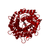 8h1kC 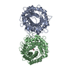 8h1lC  8h1mC 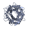 3vw5S S: Starting model for refinement C: citing same article ( |
|---|---|
| Similar structure data |
-Links
 PDBj
PDBj
- Assembly
| Deposited unit | 
| ||||||||||||
|---|---|---|---|---|---|---|---|---|---|---|---|---|---|
| 1 | 
| ||||||||||||
| Unit cell |
|
-Components
| #1: Protein | Mass: 49039.062 Da / Num. of mol.: 1 / Mutation: D254A Source method: isolated from a genetically manipulated source Source: (gene. exp.) Runella slithyformis (bacteria) / Gene: Runsl_4512 / Production host: | ||||
|---|---|---|---|---|---|
| #2: Sugar | ChemComp-SOR / 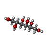  Type: D-saccharide / Mass: 182.172 Da / Num. of mol.: 1 / Source method: obtained synthetically / Formula: C6H14O6 / Feature type: SUBJECT OF INVESTIGATION Type: D-saccharide / Mass: 182.172 Da / Num. of mol.: 1 / Source method: obtained synthetically / Formula: C6H14O6 / Feature type: SUBJECT OF INVESTIGATION | ||||
| #3: Chemical |   Mass: 46.025 Da / Num. of mol.: 3 / Source method: isolated from a natural source / Formula: CH2O2 Mass: 46.025 Da / Num. of mol.: 3 / Source method: isolated from a natural source / Formula: CH2O2#4: Water | ChemComp-HOH / |  Mass: 18.015 Da / Num. of mol.: 117 / Source method: isolated from a natural source / Formula: H2O Mass: 18.015 Da / Num. of mol.: 117 / Source method: isolated from a natural source / Formula: H2OHas ligand of interest | Y | |
-Experimental details
-Experiment
| Experiment | Method: X-RAY DIFFRACTION / Number of used crystals: 1 |
|---|
- Sample preparation
| Crystal | Density Matthews: 3.82 Å3/Da / Density % sol: 67.83 % |
|---|---|
| Crystal grow | Temperature: 293.15 K / Method: vapor diffusion, sitting drop / Details: 3.6M sodium formate, 486 mM D-glucitol |
-Data collection
| Diffraction | Mean temperature: 80 K / Serial crystal experiment: N |
|---|---|
| Diffraction source | Source: SYNCHROTRON / Site: SPring-8 / Beamline: BL45XU / Wavelength: 1 Å |
| Detector | Type: DECTRIS PILATUS 6M / Detector: PIXEL / Date: May 21, 2022 |
| Radiation | Protocol: SINGLE WAVELENGTH / Monochromatic (M) / Laue (L): M / Scattering type: x-ray |
| Radiation wavelength | Wavelength: 1 Å / Relative weight: 1 |
| Reflection | Resolution: 2.67→46.08 Å / Num. obs: 20958 / % possible obs: 99.69 % / Redundancy: 25.9 % / Biso Wilson estimate: 35.21 Å2 / CC1/2: 0.996 / Rmerge(I) obs: 0.69 / Net I/σ(I): 8.68 |
| Reflection shell | Resolution: 2.67→2.77 Å / Num. unique obs: 1995 / CC1/2: 0.675 |
- Processing
| Software |
| ||||||||||||||||||||||||||||||||||||||||||||||||||||||||
|---|---|---|---|---|---|---|---|---|---|---|---|---|---|---|---|---|---|---|---|---|---|---|---|---|---|---|---|---|---|---|---|---|---|---|---|---|---|---|---|---|---|---|---|---|---|---|---|---|---|---|---|---|---|---|---|---|---|
| Refinement | Method to determine structure: MOLECULAR REPLACEMENT Starting model: 3vw5 Resolution: 2.67→46.08 Å / SU ML: 0.335 / Cross valid method: FREE R-VALUE / σ(F): 1.34 / Phase error: 25.5066 Stereochemistry target values: GeoStd + Monomer Library + CDL v1.2
| ||||||||||||||||||||||||||||||||||||||||||||||||||||||||
| Solvent computation | Shrinkage radii: 0.9 Å / VDW probe radii: 1.11 Å / Solvent model: FLAT BULK SOLVENT MODEL | ||||||||||||||||||||||||||||||||||||||||||||||||||||||||
| Displacement parameters | Biso mean: 38.94 Å2 | ||||||||||||||||||||||||||||||||||||||||||||||||||||||||
| Refinement step | Cycle: LAST / Resolution: 2.67→46.08 Å
| ||||||||||||||||||||||||||||||||||||||||||||||||||||||||
| Refine LS restraints |
| ||||||||||||||||||||||||||||||||||||||||||||||||||||||||
| LS refinement shell |
|
