 Movie
Movie Controller

[English] 日本語
 Yorodumi
- PDB-8gtt: Cryo-EM structure of human Pannexin1 resembling Pannexin2 pore wi... -
+ Open data

- Basic information
| Entry | Database: PDB / ID: 8gtt | ||||||
|---|---|---|---|---|---|---|---|
| Title | Cryo-EM structure of human Pannexin1 resembling Pannexin2 pore with W74R/R75Dmutations | ||||||
 Components Components | Pannexin-1 | ||||||
 Keywords Keywords | MEMBRANE PROTEIN / Pannexin1 / ATP release / large-pore ion channel. | ||||||
| Function / homology |  Function and homology information Function and homology informationATP transmembrane transporter activity / ATP transport / leak channel activity / Electric Transmission Across Gap Junctions / positive regulation of interleukin-1 alpha production / monoatomic anion transmembrane transport / wide pore channel activity / bleb / monoatomic anion channel activity / gap junction ...ATP transmembrane transporter activity / ATP transport / leak channel activity / Electric Transmission Across Gap Junctions / positive regulation of interleukin-1 alpha production / monoatomic anion transmembrane transport / wide pore channel activity / bleb / monoatomic anion channel activity / gap junction / gap junction channel activity / positive regulation of macrophage cytokine production / oogenesis / Mechanical load activates signaling by PIEZO1 and integrins in osteocytes / response to ATP / The NLRP3 inflammasome / monoatomic cation transport / response to ischemia / positive regulation of interleukin-1 beta production / calcium channel activity / calcium ion transport / actin filament binding / cell-cell signaling / protease binding / scaffold protein binding / High laminar flow shear stress activates signaling by PIEZO1 and PECAM1:CDH5:KDR in endothelial cells / transmembrane transporter binding / signaling receptor binding / endoplasmic reticulum membrane / structural molecule activity / endoplasmic reticulum / protein-containing complex / membrane / identical protein binding / plasma membrane Similarity search - Function | ||||||
| Biological species |  Homo sapiens (human) Homo sapiens (human) | ||||||
| Method | ELECTRON MICROSCOPY / single particle reconstruction / cryo EM / Resolution: 3.2 Å | ||||||
 Authors Authors | Hussain, N. / Penmatsa, A. | ||||||
| Funding support |  India, 1items India, 1items
| ||||||
 Citation Citation | Journal: Nat Commun / Year: 2024 Title: Cryo-EM structures of pannexin 1 and 3 reveal differences among pannexin isoforms. Authors: Nazia Hussain / Ashish Apotikar / Shabareesh Pidathala / Sourajit Mukherjee / Ananth Prasad Burada / Sujit Kumar Sikdar / Kutti R Vinothkumar / Aravind Penmatsa /  Abstract: Pannexins are single-membrane large-pore channels that release ions and ATP upon activation. Three isoforms of pannexins 1, 2, and 3, perform diverse cellular roles and differ in their pore lining ...Pannexins are single-membrane large-pore channels that release ions and ATP upon activation. Three isoforms of pannexins 1, 2, and 3, perform diverse cellular roles and differ in their pore lining residues. In this study, we report the cryo-EM structure of pannexin 3 at 3.9 Å and analyze its structural differences with pannexin isoforms 1 and 2. The pannexin 3 vestibule has two distinct chambers and a wider pore radius in comparison to pannexins 1 and 2. We further report two cryo-EM structures of pannexin 1, with pore substitutions W74R/R75D that mimic the pore lining residues of pannexin 2 and a germline mutant of pannexin 1, R217H at resolutions of 3.2 Å and 3.9 Å, respectively. Substitution of cationic residues in the vestibule of pannexin 1 results in reduced ATP interaction propensities to the channel. The germline mutant R217H in transmembrane helix 3 (TM3), leads to a partially constricted pore, reduced ATP interaction and weakened voltage sensitivity. The study compares the three pannexin isoform structures, the effects of substitutions of pore and vestibule-lining residues and allosteric effects of a pathological substitution on channel structure and function thereby enhancing our understanding of this vital group of ATP-release channels. | ||||||
| History |
|
- Structure visualization
| Structure viewer | Molecule: MolmilJmol/JSmol |
|---|
- Downloads & links
-Download
| PDBx/mmCIF format | 8gtt.cif.gz | 405.3 KB | Display | PDBx/mmCIF format |
|---|---|---|---|---|
| PDB format | pdb8gtt.ent.gz | 327.4 KB | Display | PDB format |
| PDBx/mmJSON format | 8gtt.json.gz | Tree view | PDBx/mmJSON format | |
| Others |  Other downloads Other downloads |
-Validation report
| Arichive directory | https://data.pdbj.org/pub/pdb/validation_reports/gt/8gttftp://data.pdbj.org/pub/pdb/validation_reports/gt/8gtt | HTTPS FTP |
|---|
-Related structure data
| Related structure data |  34267MC 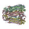 8gtrC  8gtsC M: map data used to model this data C: citing same article ( |
|---|---|
| Similar structure data |


-Links
 PDBj
PDBj


- Assembly
| Deposited unit | 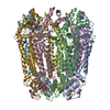
|
|---|---|
| 1 |
|
-Components
| #1: Protein | Mass: 49127.902 Da / Num. of mol.: 7 / Mutation: W74R, R75D Source method: isolated from a genetically manipulated source Source: (gene. exp.) Homo sapiens (human) / Gene: PANX1, MRS1, UNQ2529/PRO6028 / Cell line (production host): HEK293S / Production host: Homo sapiens (human) / References: UniProt: Q96RD7Has protein modification | Y | |
|---|
-Experimental details
-Experiment
| Experiment | Method: ELECTRON MICROSCOPY |
|---|---|
| EM experiment | Aggregation state: PARTICLE / 3D reconstruction method: single particle reconstruction |
- Sample preparation
| Component | Name: Pannexin 1 (W74R/R75D) / Type: COMPLEX Details: human isoform 1 of Pannexin. Expressed in plasma membranes involved in ATP release Entity ID: all / Source: RECOMBINANT | |||||||||||||||||||||||||
|---|---|---|---|---|---|---|---|---|---|---|---|---|---|---|---|---|---|---|---|---|---|---|---|---|---|---|
| Molecular weight | Value: 48 kDa/nm / Experimental value: NO | |||||||||||||||||||||||||
| Source (natural) | Organism: Homo sapiens (human) | |||||||||||||||||||||||||
| Source (recombinant) | Organism: Homo sapiens (human) / Cell: HEK293S / Plasmid: pEG Bacmam | |||||||||||||||||||||||||
| Buffer solution | pH: 8 Details: Fresh solution containing detergent was prepared for every prep. | |||||||||||||||||||||||||
| Buffer component |
| |||||||||||||||||||||||||
| Specimen | Conc.: 6 mg/ml / Embedding applied: NO / Shadowing applied: NO / Staining applied: NO / Vitrification applied: YES / Details: Sample is homoheptamer purified to homogeneity. | |||||||||||||||||||||||||
| Specimen support | Grid material: GOLD / Grid mesh size: 300 divisions/in. / Grid type: Quantifoil R1.2/1.3 | |||||||||||||||||||||||||
| Vitrification | Instrument: FEI VITROBOT MARK IV / Cryogen name: ETHANE / Humidity: 100 % / Chamber temperature: 288 K / Details: 3.5 s single blot |
- Electron microscopy imaging
| Experimental equipment |  Model: Titan Krios / Image courtesy: FEI Company |
|---|---|
| Microscopy | Model: FEI TITAN KRIOS |
| Electron gun | Electron source:  FIELD EMISSION GUN / Accelerating voltage: 300 kV / Illumination mode: OTHER FIELD EMISSION GUN / Accelerating voltage: 300 kV / Illumination mode: OTHER |
| Electron lens | Mode: OTHER / Nominal magnification: 105000 X / Nominal defocus max: 2400 nm / Nominal defocus min: 1000 nm / Cs: 2.7 mm / Alignment procedure: COMA FREE |
| Specimen holder | Cryogen: NITROGEN / Specimen holder model: FEI TITAN KRIOS AUTOGRID HOLDER / Temperature (max): 100 K / Temperature (min): 100 K |
| Image recording | Electron dose: 39.84 e/Å2 / Detector mode: COUNTING / Film or detector model: GATAN K3 BIOQUANTUM (6k x 4k) / Num. of grids imaged: 1 / Num. of real images: 15550 |
| EM imaging optics | Energyfilter name: GIF Bioquantum / Chromatic aberration corrector: None / Details: Gatan k3 / Energyfilter slit width: 20 eV / Phase plate: OTHER / Spherical aberration corrector: None |
| Image scans | Movie frames/image: 40 / Used frames/image: 1-40 |
- Processing
| EM software |
| ||||||||||||||||||||||||||||||||||||||||
|---|---|---|---|---|---|---|---|---|---|---|---|---|---|---|---|---|---|---|---|---|---|---|---|---|---|---|---|---|---|---|---|---|---|---|---|---|---|---|---|---|---|
| Image processing | Details: Images were screened for ice thickness | ||||||||||||||||||||||||||||||||||||||||
| CTF correction | Type: PHASE FLIPPING AND AMPLITUDE CORRECTION | ||||||||||||||||||||||||||||||||||||||||
| Particle selection | Num. of particles selected: 3869579 / Details: selected through particle picking | ||||||||||||||||||||||||||||||||||||||||
| Symmetry | Point symmetry: C7 (7 fold cyclic) | ||||||||||||||||||||||||||||||||||||||||
| 3D reconstruction | Resolution: 3.2 Å / Resolution method: FSC 0.143 CUT-OFF / Num. of particles: 80029 / Num. of class averages: 23 / Symmetry type: POINT | ||||||||||||||||||||||||||||||||||||||||
| Atomic model building | B value: 106.6 / Protocol: AB INITIO MODEL / Space: REAL / Target criteria: Correlation coeficient | ||||||||||||||||||||||||||||||||||||||||
| Atomic model building | PDB-ID: 6WBF Pdb chain-ID: A / Accession code: 6WBF / Source name: PDB / Type: experimental model |

