Movie
Movie Controller

[English] 日本語
 Yorodumi
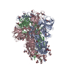
- PDB-8gs6: Structure of the SARS-CoV-2 BA.2.75 spike glycoprotein (closed st... -
+ Open data

- Basic information
| Entry | Database: PDB / ID: 8gs6 | |||||||||||||||||||||
|---|---|---|---|---|---|---|---|---|---|---|---|---|---|---|---|---|---|---|---|---|---|---|
| Title | Structure of the SARS-CoV-2 BA.2.75 spike glycoprotein (closed state 1) | |||||||||||||||||||||
 Components Components | Spike glycoprotein | |||||||||||||||||||||
 Keywords Keywords | VIRAL PROTEIN / spike glycoprotein | |||||||||||||||||||||
| Function / homology |  Function and homology information Function and homology informationsymbiont-mediated disruption of host tissue / Maturation of spike protein / Translation of Structural Proteins / Virion Assembly and Release / host cell surface / host extracellular region / symbiont-mediated-mediated suppression of host tetherin activity / Induction of Cell-Cell Fusion / structural constituent of virion / positive regulation of viral entry into host cell ...symbiont-mediated disruption of host tissue / Maturation of spike protein / Translation of Structural Proteins / Virion Assembly and Release / host cell surface / host extracellular region / symbiont-mediated-mediated suppression of host tetherin activity / Induction of Cell-Cell Fusion / structural constituent of virion / positive regulation of viral entry into host cell / membrane fusion / host cell endoplasmic reticulum-Golgi intermediate compartment membrane / Attachment and Entry / entry receptor-mediated virion attachment to host cell / receptor-mediated virion attachment to host cell / host cell surface receptor binding / symbiont-mediated suppression of host innate immune response / endocytosis involved in viral entry into host cell / receptor ligand activity / fusion of virus membrane with host plasma membrane / fusion of virus membrane with host endosome membrane / viral envelope / symbiont entry into host cell / virion attachment to host cell / host cell plasma membrane / SARS-CoV-2 activates/modulates innate and adaptive immune responses / virion membrane / membrane / identical protein binding / plasma membrane Similarity search - Function | |||||||||||||||||||||
| Biological species |   Severe acute respiratory syndrome coronavirus 2 Severe acute respiratory syndrome coronavirus 2 | |||||||||||||||||||||
| Method | ELECTRON MICROSCOPY / single particle reconstruction / cryo EM / Resolution: 2.86 Å | |||||||||||||||||||||
 Authors Authors | Anraku, Y. / Tabata-Sasaki, K. / Kita, S. / Fukuhara, H. / Maenaka, K. / Hashiguchi, T. | |||||||||||||||||||||
| Funding support |  Japan, 6items Japan, 6items
| |||||||||||||||||||||
 Citation Citation | Journal: Cell Host Microbe / Year: 2022 Title: Virological characteristics of the SARS-CoV-2 Omicron BA.2.75 variant. Authors: Akatsuki Saito / Tomokazu Tamura / Jiri Zahradnik / Sayaka Deguchi / Koshiro Tabata / Yuki Anraku / Izumi Kimura / Jumpei Ito / Daichi Yamasoba / Hesham Nasser / Mako Toyoda / Kayoko Nagata ...Authors: Akatsuki Saito / Tomokazu Tamura / Jiri Zahradnik / Sayaka Deguchi / Koshiro Tabata / Yuki Anraku / Izumi Kimura / Jumpei Ito / Daichi Yamasoba / Hesham Nasser / Mako Toyoda / Kayoko Nagata / Keiya Uriu / Yusuke Kosugi / Shigeru Fujita / Maya Shofa / Mst Monira Begum / Ryo Shimizu / Yoshitaka Oda / Rigel Suzuki / Hayato Ito / Naganori Nao / Lei Wang / Masumi Tsuda / Kumiko Yoshimatsu / Jin Kuramochi / Shunsuke Kita / Kaori Sasaki-Tabata / Hideo Fukuhara / Katsumi Maenaka / Yuki Yamamoto / Tetsuharu Nagamoto / Hiroyuki Asakura / Mami Nagashima / Kenji Sadamasu / Kazuhisa Yoshimura / Takamasa Ueno / Gideon Schreiber / Akifumi Takaori-Kondo / / Kotaro Shirakawa / Hirofumi Sawa / Takashi Irie / Takao Hashiguchi / Kazuo Takayama / Keita Matsuno / Shinya Tanaka / Terumasa Ikeda / Takasuke Fukuhara / Kei Sato /   Abstract: The SARS-CoV-2 Omicron BA.2.75 variant emerged in May 2022. BA.2.75 is a BA.2 descendant but is phylogenetically distinct from BA.5, the currently predominant BA.2 descendant. Here, we show that BA.2. ...The SARS-CoV-2 Omicron BA.2.75 variant emerged in May 2022. BA.2.75 is a BA.2 descendant but is phylogenetically distinct from BA.5, the currently predominant BA.2 descendant. Here, we show that BA.2.75 has a greater effective reproduction number and different immunogenicity profile than BA.5. We determined the sensitivity of BA.2.75 to vaccinee and convalescent sera as well as a panel of clinically available antiviral drugs and antibodies. Antiviral drugs largely retained potency, but antibody sensitivity varied depending on several key BA.2.75-specific substitutions. The BA.2.75 spike exhibited a profoundly higher affinity for its human receptor, ACE2. Additionally, the fusogenicity, growth efficiency in human alveolar epithelial cells, and intrinsic pathogenicity in hamsters of BA.2.75 were greater than those of BA.2. Our multilevel investigations suggest that BA.2.75 acquired virological properties independent of BA.5, and the potential risk of BA.2.75 to global health is greater than that of BA.5. | |||||||||||||||||||||
| History |
|
- Structure visualization
| Structure viewer | Molecule: MolmilJmol/JSmol |
|---|
- Downloads & links
-Download
| PDBx/mmCIF format | 8gs6.cif.gz | 577.2 KB | Display | PDBx/mmCIF format |
|---|---|---|---|---|
| PDB format | pdb8gs6.ent.gz | 450.4 KB | Display | PDB format |
| PDBx/mmJSON format | 8gs6.json.gz | Tree view | PDBx/mmJSON format | |
| Others |  Other downloads Other downloads |
-Validation report
| Arichive directory | https://data.pdbj.org/pub/pdb/validation_reports/gs/8gs6ftp://data.pdbj.org/pub/pdb/validation_reports/gs/8gs6 | HTTPS FTP |
|---|
-Related structure data
| Related structure data | 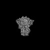 34221MC M: map data used to model this data C: citing same article ( |
|---|---|
| Similar structure data |


-Links
 PDBj
PDBj


- Assembly
| Deposited unit | 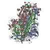
|
|---|---|
| 1 |
|
-Components
| #1: Protein | Mass: 138332.453 Da / Num. of mol.: 3 Mutation: R682G, R683S, R685G, F817P, A892P, A899P, A942P, K986P, V987P Source method: isolated from a genetically manipulated source Source: (gene. exp.) Severe acute respiratory syndrome coronavirus 2Strain: BA.2.75 / Gene: S, 2 / Cell line (production host): HEK293 / Production host:  Homo sapiens (human) / References: UniProt: P0DTC2 Homo sapiens (human) / References: UniProt: P0DTC2#2: Polysaccharide | 2-acetamido-2-deoxy-beta-D-glucopyranose-(1-4)-2-acetamido-2-deoxy-beta-D-glucopyranose Source method: isolated from a genetically manipulated source #3: Sugar | ChemComp-NAG /   Type: D-saccharide, beta linking / Mass: 221.208 Da / Num. of mol.: 31 / Source method: obtained synthetically / Formula: C8H15NO6 Type: D-saccharide, beta linking / Mass: 221.208 Da / Num. of mol.: 31 / Source method: obtained synthetically / Formula: C8H15NO6Has ligand of interest | N | Has protein modification | Y | |
|---|
-Experimental details
-Experiment
| Experiment | Method: ELECTRON MICROSCOPY |
|---|---|
| EM experiment | Aggregation state: PARTICLE / 3D reconstruction method: single particle reconstruction |
- Sample preparation
| Component | Name: SARS-COV-2 BA.2.75 spike glycoprotein / Type: COMPLEX / Entity ID: #1 / Source: RECOMBINANT |
|---|---|
| Molecular weight | Value: 0.42 MDa / Experimental value: NO |
| Source (natural) | Organism: Severe acute respiratory syndrome coronavirus 2 |
| Source (recombinant) | Organism: Homo sapiens (human) |
| Buffer solution | pH: 7.4 Details: Octyl glucoside solution was added to PBS solution to a final concentration of 0.01% |
| Specimen | Conc.: 1.2 mg/ml / Embedding applied: NO / Shadowing applied: NO / Staining applied: NO / Vitrification applied: YES |
| Specimen support | Grid material: COPPER / Grid mesh size: 300 divisions/in. / Grid type: Quantifoil R2/2 |
| Vitrification | Instrument: FEI VITROBOT MARK IV / Cryogen name: ETHANE / Humidity: 100 % / Chamber temperature: 291 K / Details: blotting time 5 s and blotting force 5. |
- Electron microscopy imaging
| Experimental equipment |  Model: Titan Krios / Image courtesy: FEI Company |
|---|---|
| Microscopy | Model: TFS KRIOS |
| Electron gun | Electron source:  FIELD EMISSION GUN / Accelerating voltage: 300 kV / Illumination mode: FLOOD BEAM FIELD EMISSION GUN / Accelerating voltage: 300 kV / Illumination mode: FLOOD BEAM |
| Electron lens | Mode: BRIGHT FIELD / Nominal magnification: 130000 X / Nominal defocus max: 1800 nm / Nominal defocus min: 800 nm / Cs: 2.7 mm |
| Specimen holder | Cryogen: NITROGEN / Specimen holder model: FEI TITAN KRIOS AUTOGRID HOLDER |
| Image recording | Average exposure time: 1.5 sec. / Electron dose: 50 e/Å2 / Film or detector model: GATAN K3 BIOQUANTUM (6k x 4k) / Num. of real images: 3308 |
| EM imaging optics | Energyfilter name: GIF Bioquantum / Energyfilter slit width: 20 eV |
| Image scans | Width: 5760 / Height: 4092 |
- Processing
| Software |
| ||||||||||||||||||||||||||||||||||||||||
|---|---|---|---|---|---|---|---|---|---|---|---|---|---|---|---|---|---|---|---|---|---|---|---|---|---|---|---|---|---|---|---|---|---|---|---|---|---|---|---|---|---|
| EM software |
| ||||||||||||||||||||||||||||||||||||||||
| CTF correction | Type: PHASE FLIPPING AND AMPLITUDE CORRECTION | ||||||||||||||||||||||||||||||||||||||||
| Particle selection | Num. of particles selected: 754589 | ||||||||||||||||||||||||||||||||||||||||
| Symmetry | Point symmetry: C1 (asymmetric) | ||||||||||||||||||||||||||||||||||||||||
| 3D reconstruction | Resolution: 2.86 Å / Resolution method: FSC 0.143 CUT-OFF / Num. of particles: 139418 / Algorithm: FOURIER SPACE / Num. of class averages: 1 / Symmetry type: POINT | ||||||||||||||||||||||||||||||||||||||||
| Atomic model building | Protocol: RIGID BODY FIT / Space: REAL | ||||||||||||||||||||||||||||||||||||||||
| Atomic model building | PDB-ID: 7UB0 Accession code: 7UB0 / Source name: PDB / Type: experimental model | ||||||||||||||||||||||||||||||||||||||||
| Refinement | Cross valid method: NONE Stereochemistry target values: GeoStd + Monomer Library + CDL v1.2 | ||||||||||||||||||||||||||||||||||||||||
| Displacement parameters | Biso mean: 90.1 Å2 | ||||||||||||||||||||||||||||||||||||||||
| Refine LS restraints |
|