 Movie
Movie Controller

[English] 日本語
 Yorodumi
- PDB-8gr6: Crystal Structure of pilus-specific Sortase C from Streptococcus ... -
+ Open data

- Basic information
| Entry | Database: PDB / ID: 8gr6 | ||||||
|---|---|---|---|---|---|---|---|
| Title | Crystal Structure of pilus-specific Sortase C from Streptococcus sanguinis | ||||||
 Components Components | Sortase-like protein, putative | ||||||
 Keywords Keywords | HYDROLASE / Sortase C / Pilus specific sortase / Cysteine-transpeptidase / Streptococcus sanguinis / dental plaque / biofilm | ||||||
| Function / homology | Sortase C / Sortase family / Sortase domain superfamily / Sortase domain / metal ion binding / membrane / Sortase-like protein, putative Function and homology information Function and homology information | ||||||
| Biological species |  Streptococcus sanguinis SK36 (bacteria) Streptococcus sanguinis SK36 (bacteria) | ||||||
| Method |  X-RAY DIFFRACTION / SYNCHROTRON / MOLECULAR REPLACEMENT / Resolution: 2.06 Å X-RAY DIFFRACTION / SYNCHROTRON / MOLECULAR REPLACEMENT / Resolution: 2.06 Å | ||||||
 Authors Authors | Yadav, S. / Parijat, P. / Krishnan, V. | ||||||
| Funding support |  India, 1items India, 1items
| ||||||
 Citation Citation | Journal: Int.J.Biol.Macromol. / Year: 2023 Title: Crystal structure of the pilus-specific sortase from early colonizing oral Streptococcus sanguinis captures an active open-lid conformation. Authors: Yadav, S. / Parijat, P. / Krishnan, V. | ||||||
| History |
|
- Structure visualization
| Structure viewer | Molecule: MolmilJmol/JSmol |
|---|
- Downloads & links
-Download
| PDBx/mmCIF format | 8gr6.cif.gz | 100.3 KB | Display | PDBx/mmCIF format |
|---|---|---|---|---|
| PDB format | pdb8gr6.ent.gz | 74 KB | Display | PDB format |
| PDBx/mmJSON format | 8gr6.json.gz | Tree view | PDBx/mmJSON format | |
| Others |  Other downloads Other downloads |
-Validation report
| Summary document | 8gr6_validation.pdf.gz | 442.1 KB | Display | wwPDB validaton report |
|---|---|---|---|---|
| Full document | 8gr6_full_validation.pdf.gz | 441.9 KB | Display | |
| Data in XML | 8gr6_validation.xml.gz | 10.3 KB | Display | |
| Data in CIF | 8gr6_validation.cif.gz | 13.7 KB | Display | |
| Arichive directory | https://data.pdbj.org/pub/pdb/validation_reports/gr/8gr6ftp://data.pdbj.org/pub/pdb/validation_reports/gr/8gr6 | HTTPS FTP |
-Related structure data
| Related structure data |  8guuC  4d7wS S: Starting model for refinement C: citing same article ( |
|---|---|
| Similar structure data |
-Links
 PDBj
PDBj- Assembly
| Deposited unit | 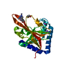
| |||||||||
|---|---|---|---|---|---|---|---|---|---|---|
| 1 |
| |||||||||
| Unit cell |
| |||||||||
| Components on special symmetry positions |
|
-Components
| #1: Protein | Mass: 24005.969 Da / Num. of mol.: 1 Source method: isolated from a genetically manipulated source Source: (gene. exp.) Streptococcus sanguinis SK36 (bacteria)Strain: SK36 / Gene: srtC, SSA_1631 / Variant: No / Plasmid: pET28b / Production host: | ||||
|---|---|---|---|---|---|
| #2: Chemical | ChemComp-EDO /   Mass: 62.068 Da / Num. of mol.: 1 / Source method: obtained synthetically / Formula: C2H6O2 Mass: 62.068 Da / Num. of mol.: 1 / Source method: obtained synthetically / Formula: C2H6O2 | ||||
| #3: Chemical | ChemComp-ZN /   Mass: 65.409 Da / Num. of mol.: 1 / Source method: obtained synthetically / Formula: Zn Mass: 65.409 Da / Num. of mol.: 1 / Source method: obtained synthetically / Formula: Zn | ||||
| #4: Chemical |   Mass: 22.990 Da / Num. of mol.: 2 / Source method: obtained synthetically / Formula: Na Mass: 22.990 Da / Num. of mol.: 2 / Source method: obtained synthetically / Formula: Na#5: Water | ChemComp-HOH / |  Mass: 18.015 Da / Num. of mol.: 76 / Source method: isolated from a natural source / Formula: H2O Mass: 18.015 Da / Num. of mol.: 76 / Source method: isolated from a natural source / Formula: H2OHas ligand of interest | N | |
-Experimental details
-Experiment
| Experiment | Method: X-RAY DIFFRACTION / Number of used crystals: 1 |
|---|
- Sample preparation
| Crystal | Density Matthews: 4.05 Å3/Da / Density % sol: 69 % / Description: Rhombohedral |
|---|---|
| Crystal grow | Temperature: 295 K / Method: vapor diffusion, hanging drop / pH: 5.5 Details: 100mM Sodium acetate (pH 5.5), 200mM Ammonium-Sulphate and 10% (w/v) PEG 2000 MME and 12.5 mM Zinc-Chloride |
-Data collection
| Diffraction | Mean temperature: 100 K / Serial crystal experiment: N |
|---|---|
| Diffraction source | Source: SYNCHROTRON / Site: ESRF  / Beamline: MASSIF-1 / Wavelength: 0.965459 Å / Beamline: MASSIF-1 / Wavelength: 0.965459 Å |
| Detector | Type: DECTRIS PILATUS3 2M / Detector: PIXEL / Date: Mar 4, 2021 |
| Radiation | Protocol: SINGLE WAVELENGTH / Monochromatic (M) / Laue (L): M / Scattering type: x-ray |
| Radiation wavelength | Wavelength: 0.965459 Å / Relative weight: 1 |
| Reflection | Resolution: 2.056→52.331 Å / Num. obs: 23670 / % possible obs: 97.2 % / Redundancy: 6.6 % / CC1/2: 0.999 / Rmerge(I) obs: 0.06 / Rpim(I) all: 0.024 / Rrim(I) all: 0.065 / Net I/σ(I): 13.7 |
| Reflection shell | Resolution: 2.056→2.092 Å / Redundancy: 7.1 % / Rmerge(I) obs: 0.58 / Mean I/σ(I) obs: 2.4 / Num. unique obs: 1220 / CC1/2: 0.91 / Rpim(I) all: 0.23 / Rrim(I) all: 0.625 / % possible all: 100 |
- Processing
| Software |
| |||||||||||||||||||||||||||||||||||||||||||||||||||||||||||||||||||||||||||||||||||||||||||||||||||||||||
|---|---|---|---|---|---|---|---|---|---|---|---|---|---|---|---|---|---|---|---|---|---|---|---|---|---|---|---|---|---|---|---|---|---|---|---|---|---|---|---|---|---|---|---|---|---|---|---|---|---|---|---|---|---|---|---|---|---|---|---|---|---|---|---|---|---|---|---|---|---|---|---|---|---|---|---|---|---|---|---|---|---|---|---|---|---|---|---|---|---|---|---|---|---|---|---|---|---|---|---|---|---|---|---|---|---|---|
| Refinement | Method to determine structure: MOLECULAR REPLACEMENT Starting model: 4D7W Resolution: 2.06→52.33 Å / Cor.coef. Fo:Fc: 0.968 / Cor.coef. Fo:Fc free: 0.964 / SU B: 7.232 / SU ML: 0.093 / Cross valid method: THROUGHOUT / ESU R: 0.122 / ESU R Free: 0.114 / Stereochemistry target values: MAXIMUM LIKELIHOOD Details: U VALUES : WITH TLS ADDED HYDROGENS HAVE BEEN ADDED IN THE RIDING POSITIONS U VALUES : RESIDUAL ONLY
| |||||||||||||||||||||||||||||||||||||||||||||||||||||||||||||||||||||||||||||||||||||||||||||||||||||||||
| Solvent computation | Ion probe radii: 0.7 Å / Shrinkage radii: 0.7 Å / VDW probe radii: 1.1 Å / Solvent model: MASK | |||||||||||||||||||||||||||||||||||||||||||||||||||||||||||||||||||||||||||||||||||||||||||||||||||||||||
| Displacement parameters | Biso mean: 56.162 Å2
| |||||||||||||||||||||||||||||||||||||||||||||||||||||||||||||||||||||||||||||||||||||||||||||||||||||||||
| Refinement step | Cycle: LAST / Resolution: 2.06→52.33 Å
| |||||||||||||||||||||||||||||||||||||||||||||||||||||||||||||||||||||||||||||||||||||||||||||||||||||||||
| Refine LS restraints |
| |||||||||||||||||||||||||||||||||||||||||||||||||||||||||||||||||||||||||||||||||||||||||||||||||||||||||
| LS refinement shell | Resolution: 2.06→2.11 Å
| |||||||||||||||||||||||||||||||||||||||||||||||||||||||||||||||||||||||||||||||||||||||||||||||||||||||||
| Refinement TLS params. | Method: refined / Origin x: 14.694 Å / Origin y: -15.999 Å / Origin z: -16.597 Å
|
