 ムービー
ムービー コントローラー


+ データを開く

- 基本情報
基本情報
| 登録情報 | データベース: PDB / ID: 8guu | ||||||
|---|---|---|---|---|---|---|---|
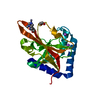
| タイトル | Crystal structure of pilus-specific sortase C mutant from Streptococcus sanguinis | ||||||
 要素 要素 | Sortase-like protein, putative | ||||||
 キーワード キーワード | HYDROLASE / Sortase C / pilus-specific sortase / Cysteine transpeptidase / dental plaque / biofilm / pili | ||||||
| 機能・相同性 | Sortase C / Sortase family / Sortase domain superfamily / Sortase domain / metal ion binding / membrane / Sortase-like protein, putative 機能・相同性情報 機能・相同性情報 | ||||||
| 生物種 |  Streptococcus sanguinis SK36 (バクテリア) Streptococcus sanguinis SK36 (バクテリア) | ||||||
| 手法 |  X線回折 / シンクロトロン / 分子置換 / 解像度: 2.008 Å X線回折 / シンクロトロン / 分子置換 / 解像度: 2.008 Å | ||||||
 データ登録者 データ登録者 | Yadav, S. / Parijat, P. / Krishnan, V. | ||||||
| 資金援助 |  インド, 1件 インド, 1件
| ||||||
 引用 引用 | ジャーナル: Int.J.Biol.Macromol. / 年: 2023 タイトル: Crystal structure of the pilus-specific sortase from early colonizing oral Streptococcus sanguinis captures an active open-lid conformation. 著者: Yadav, S. / Parijat, P. / Krishnan, V. | ||||||
| 履歴 |
|
- 構造の表示
構造の表示
| 構造ビューア | 分子: MolmilJmol/JSmol |
|---|
- ダウンロードとリンク
-ダウンロード
| PDBx/mmCIF形式 | 8guu.cif.gz | 95.6 KB | 表示 | PDBx/mmCIF形式 |
|---|---|---|---|---|
| PDB形式 | pdb8guu.ent.gz | 71.2 KB | 表示 | PDB形式 |
| PDBx/mmJSON形式 | 8guu.json.gz | ツリー表示 | PDBx/mmJSON形式 | |
| その他 |  その他のダウンロード その他のダウンロード |
-検証レポート
| アーカイブディレクトリ | https://data.pdbj.org/pub/pdb/validation_reports/gu/8guuftp://data.pdbj.org/pub/pdb/validation_reports/gu/8guu | HTTPS FTP |
|---|
-関連構造データ
-リンク
 PDBj
PDBj- 集合体
集合体
| 登録構造単位 | 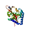
| ||||||||
|---|---|---|---|---|---|---|---|---|---|
| 1 |
| ||||||||
| 単位格子 |
| ||||||||
| Components on special symmetry positions |
|
-要素
| #1: タンパク質 | 分子量: 21688.430 Da / 分子数: 1 / 変異: C209A / 由来タイプ: 組換発現 由来: (組換発現) Streptococcus sanguinis SK36 (バクテリア)株: SK36 / 遺伝子: srtC, SSA_1631 / プラスミド: pET28b / 発現宿主: | ||||||
|---|---|---|---|---|---|---|---|
| #2: 化合物 |   分子量: 62.068 Da / 分子数: 2 / 由来タイプ: 合成 / 式: C2H6O2 / タイプ: SUBJECT OF INVESTIGATION 分子量: 62.068 Da / 分子数: 2 / 由来タイプ: 合成 / 式: C2H6O2 / タイプ: SUBJECT OF INVESTIGATION#3: 水 | ChemComp-HOH / |  分子量: 18.015 Da / 分子数: 87 / 由来タイプ: 天然 / 式: H2O 分子量: 18.015 Da / 分子数: 87 / 由来タイプ: 天然 / 式: H2O研究の焦点であるリガンドがあるか | Y | Has protein modification | Y | |
-実験情報
-実験
| 実験 | 手法: X線回折 / 使用した結晶の数: 1 |
|---|
- 試料調製
試料調製
| 結晶 | マシュー密度: 4.52 Å3/Da / 溶媒含有率: 69 % / 解説: Rhombohedral |
|---|---|
| 結晶化 | 温度: 295 K / 手法: 蒸気拡散法, ハンギングドロップ法 / pH: 5 詳細: 100 mM Sodium Acetate pH 5.5, 200 mM Ammonium sulphate, and 10 % PEG 2000MME. |
-データ収集
| 回折 | 平均測定温度: 100 K / Serial crystal experiment: N |
|---|---|
| 放射光源 | 由来: シンクロトロン / サイト: ESRF  / ビームライン: ID23-1 / 波長: 0.97242 Å / ビームライン: ID23-1 / 波長: 0.97242 Å |
| 検出器 | タイプ: DECTRIS PILATUS 6M / 検出器: PIXEL / 日付: 2020年10月30日 |
| 放射 | プロトコル: SINGLE WAVELENGTH / 単色(M)・ラウエ(L): M / 散乱光タイプ: x-ray |
| 放射波長 | 波長: 0.97242 Å / 相対比: 1 |
| 反射 | 解像度: 2.008→69.82 Å / Num. obs: 24438 / % possible obs: 93.3 % / 冗長度: 11.8 % / Biso Wilson estimate: 42.7 Å2 / CC1/2: 0.999 / Rmerge(I) obs: 0.088 / Rpim(I) all: 0.026 / Net I/σ(I): 15.9 |
| 反射 シェル | 解像度: 2.008→2.043 Å / 冗長度: 9.9 % / Rmerge(I) obs: 1.039 / Mean I/σ(I) obs: 2.2 / Num. unique obs: 12862 / CC1/2: 0.871 / Rpim(I) all: 0.348 |
- 解析
解析
| ソフトウェア |
| |||||||||||||||||||||||||||||||||||||||||||||||||||||||||||||||||||||||||||||||||||||||||||||||||||||||||
|---|---|---|---|---|---|---|---|---|---|---|---|---|---|---|---|---|---|---|---|---|---|---|---|---|---|---|---|---|---|---|---|---|---|---|---|---|---|---|---|---|---|---|---|---|---|---|---|---|---|---|---|---|---|---|---|---|---|---|---|---|---|---|---|---|---|---|---|---|---|---|---|---|---|---|---|---|---|---|---|---|---|---|---|---|---|---|---|---|---|---|---|---|---|---|---|---|---|---|---|---|---|---|---|---|---|---|
| 精密化 | 構造決定の手法: 分子置換 開始モデル: 8GR6 解像度: 2.008→69.82 Å / Cor.coef. Fo:Fc: 0.97 / Cor.coef. Fo:Fc free: 0.959 / SU B: 8.669 / SU ML: 0.111 / 交差検証法: THROUGHOUT / ESU R: 0.12 / ESU R Free: 0.119 / 立体化学のターゲット値: MAXIMUM LIKELIHOOD 詳細: U VALUES : WITH TLS ADDED HYDROGENS HAVE BEEN ADDED IN THE RIDING POSITIONS U VALUES : RESIDUAL ONLY
| |||||||||||||||||||||||||||||||||||||||||||||||||||||||||||||||||||||||||||||||||||||||||||||||||||||||||
| 溶媒の処理 | イオンプローブ半径: 0.7 Å / 減衰半径: 0.7 Å / VDWプローブ半径: 1 Å / 溶媒モデル: MASK | |||||||||||||||||||||||||||||||||||||||||||||||||||||||||||||||||||||||||||||||||||||||||||||||||||||||||
| 原子変位パラメータ | Biso mean: 54.637 Å2
| |||||||||||||||||||||||||||||||||||||||||||||||||||||||||||||||||||||||||||||||||||||||||||||||||||||||||
| 精密化ステップ | サイクル: LAST / 解像度: 2.008→69.82 Å
| |||||||||||||||||||||||||||||||||||||||||||||||||||||||||||||||||||||||||||||||||||||||||||||||||||||||||
| 拘束条件 |
| |||||||||||||||||||||||||||||||||||||||||||||||||||||||||||||||||||||||||||||||||||||||||||||||||||||||||
| LS精密化 シェル | 解像度: 2.008→2.06 Å / Total num. of bins used: 20
| |||||||||||||||||||||||||||||||||||||||||||||||||||||||||||||||||||||||||||||||||||||||||||||||||||||||||
| 精密化 TLS | 手法: refined / Origin x: 14.72 Å / Origin y: -16.008 Å / Origin z: -16.576 Å
|
