negative regulation of B cell activation / RHO GTPases Activate WASPs and WAVEs / G beta:gamma signalling through BTK / negative regulation of leukocyte proliferation / FCERI mediated Ca+2 mobilization / G alpha (12/13) signalling events / G alpha (q) signalling events / Antigen activates B Cell Receptor (BCR) leading to generation of second messengers / Regulation of actin dynamics for phagocytic cup formation / monocyte proliferation ...negative regulation of B cell activation / RHO GTPases Activate WASPs and WAVEs / G beta:gamma signalling through BTK / negative regulation of leukocyte proliferation / FCERI mediated Ca+2 mobilization / G alpha (12/13) signalling events / G alpha (q) signalling events / Antigen activates B Cell Receptor (BCR) leading to generation of second messengers / Regulation of actin dynamics for phagocytic cup formation / monocyte proliferation / positive regulation of interleukin-17A production / proteoglycan catabolic process / eosinophil homeostasis / positive regulation of type III hypersensitivity / B cell affinity maturation / positive regulation of synoviocyte proliferation / histamine secretion by mast cell / positive regulation of cGAS/STING signaling pathway / neutrophil homeostasis / positive regulation of type I hypersensitivity / cellular response to molecule of fungal origin / cellular response to interleukin-7 / DAP12 signaling / cGAS/STING signaling pathway / negative regulation of cytokine production / negative regulation of interleukin-10 production / phospholipase activator activity / negative regulation of B cell proliferation / positive regulation of immunoglobulin production / phosphatidylinositol-3,4,5-trisphosphate binding / positive regulation of NLRP3 inflammasome complex assembly / phospholipase binding / cell maturation / positive regulation of B cell proliferation / positive regulation of type I interferon production / positive regulation of phagocytosis / B cell receptor signaling pathway / non-specific protein-tyrosine kinase / cellular response to reactive oxygen species / non-membrane spanning protein tyrosine kinase activity / positive regulation of interleukin-6 production / positive regulation of tumor necrosis factor production / T cell receptor signaling pathway / protein tyrosine kinase activity / cytoplasmic vesicle / response to lipopolysaccharide / adaptive immune response / intracellular signal transduction / membrane raft / innate immune response / apoptotic process / perinuclear region of cytoplasm / zinc ion binding / ATP binding / identical protein binding / nucleus / plasma membrane / cytosol / cytoplasm Similarity search - Function
National Institutes of Health/National Institute Of Allergy and Infectious Diseases (NIH/NIAID)
United States
Citation
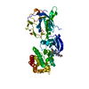
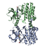
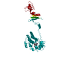
Journal: Elife / Year: 2024 Title: Conformational heterogeneity of the BTK PHTH domain drives multiple regulatory states. Authors: David Yin-Wei Lin / Lauren E Kueffer / Puneet Juneja / Thomas E Wales / John R Engen / Amy H Andreotti / Abstract: Full-length Bruton's tyrosine kinase (BTK) has been refractory to structural analysis. The nearest full-length structure of BTK to date consists of the autoinhibited SH3-SH2-kinase core. Precisely ...Full-length Bruton's tyrosine kinase (BTK) has been refractory to structural analysis. The nearest full-length structure of BTK to date consists of the autoinhibited SH3-SH2-kinase core. Precisely how the BTK N-terminal domains (the Pleckstrin homology/Tec homology [PHTH] domain and proline-rich regions [PRR] contain linker) contribute to BTK regulation remains unclear. We have produced crystals of full-length BTK for the first time but despite efforts to stabilize the autoinhibited state, the diffraction data still reveal only the SH3-SH2-kinase core with no electron density visible for the PHTH-PRR segment. Cryo-electron microscopy (cryoEM) data of full-length BTK, on the other hand, provide the first view of the PHTH domain within full-length BTK. CryoEM reconstructions support conformational heterogeneity in the PHTH-PRR region wherein the globular PHTH domain adopts a range of states arrayed around the autoinhibited SH3-SH2-kinase core. On the way to activation, disassembly of the SH3-SH2-kinase core opens a new autoinhibitory site on the kinase domain for PHTH domain binding that is ultimately released upon interaction of PHTH with phosphatidylinositol (3,4,5)-trisphosphate. Membrane-induced dimerization activates BTK and we present here a crystal structure of an activation loop swapped BTK kinase domain dimer that likely represents the conformational state leading to trans-autophosphorylation. Together, these data provide the first structural elucidation of full-length BTK and allow a deeper understanding of allosteric control over the BTK kinase domain during distinct stages of activation.
Evidence: Full-length Btk was purified as a monomer by gel-filtration. The domain-swapped dimer was formed during crystallization. The domain-swapped dimer resembles the structure 4XI2.
Mass: 664.796 Da / Num. of mol.: 1 / Source method: obtained synthetically / Formula: C37H44N8O4 / Feature type: SUBJECT OF INVESTIGATION
Has ligand of interest
Y
-
Experimental details
-
Experiment
Experiment
Method: X-RAY DIFFRACTION / Number of used crystals: 1
-
Sample preparation
Crystal
Density Matthews: 3.29 Å3/Da / Density % sol: 66.06 % Description: Monomer proteins at 18 mg/ml in 20 mM TRIS/HCl, pH 8.0, 150 mM NaCl, 10% glycerol 5 mM DTT were mixed with an equal volume of the precipitant solution, and then suspend over 0.25 ml of ...Description: Monomer proteins at 18 mg/ml in 20 mM TRIS/HCl, pH 8.0, 150 mM NaCl, 10% glycerol 5 mM DTT were mixed with an equal volume of the precipitant solution, and then suspend over 0.25 ml of precipitant solution. Leaf-shaped hexagonal crystals grew to up to 0.4 mm on a side after a period of two weeks. Crystals were frozen by transferring them, in six sequential steps, to a solution containing 0.1M Bis-Tris Propane, pH 7.5, 0.2M potassium thiocyanate, 20% PEG 3350 and 20% glycerol.
In the structure databanks used in Yorodumi, some data are registered as the other names, "COVID-19 virus" and "2019-nCoV". Here are the details of the virus and the list of structure data.
Jan 31, 2019. EMDB accession codes are about to change! (news from PDBe EMDB page)
EMDB accession codes are about to change! (news from PDBe EMDB page)
The allocation of 4 digits for EMDB accession codes will soon come to an end. Whilst these codes will remain in use, new EMDB accession codes will include an additional digit and will expand incrementally as the available range of codes is exhausted. The current 4-digit format prefixed with “EMD-” (i.e. EMD-XXXX) will advance to a 5-digit format (i.e. EMD-XXXXX), and so on. It is currently estimated that the 4-digit codes will be depleted around Spring 2019, at which point the 5-digit format will come into force.
The EM Navigator/Yorodumi systems omit the EMD- prefix.
Related info.:Q: What is EMD? / ID/Accession-code notation in Yorodumi/EM Navigator
Yorodumi is a browser for structure data from EMDB, PDB, SASBDB, etc.
This page is also the successor to EM Navigator detail page, and also detail information page/front-end page for Omokage search.
The word "yorodu" (or yorozu) is an old Japanese word meaning "ten thousand". "mi" (miru) is to see.
Related info.:EMDB / PDB / SASBDB / Comparison of 3 databanks / Yorodumi Search / Aug 31, 2016. New EM Navigator & Yorodumi / Yorodumi Papers / Jmol/JSmol / Function and homology information / Changes in new EM Navigator and Yorodumi
 Movie
Movie Controller

 Yorodumi
 Open data
 Basic information
 Components
 Keywords
 Function and homology information
 X-RAY DIFFRACTION /
  Authors
 United States, 1items
  Citation
 Structure visualization
 Downloads & links
 Other downloads


 PDBj
PDBj


 Assembly
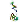


 Mass: 664.796 Da / Num. of mol.: 1 / Source method: obtained synthetically / Formula: C37H44N8O4 / Feature type: SUBJECT OF INVESTIGATION
 Sample preparation
 Processing