 Movie
Movie Controller

[English] 日本語
 Yorodumi
- PDB-8gm9: Structure of Citrate Synthase(CitA) in Mycobacterium Tuberculosis -
+ Open data

- Basic information
| Entry | Database: PDB / ID: 8gm9 | ||||||
|---|---|---|---|---|---|---|---|
| Title | Structure of Citrate Synthase(CitA) in Mycobacterium Tuberculosis | ||||||
 Components Components | citrate synthase | ||||||
 Keywords Keywords | CYTOSOLIC PROTEIN / Citrate Synthesis / TCA cycle | ||||||
| Function / homology |  Function and homology information Function and homology informationcitrate synthase (unknown stereospecificity) / citrate synthase activity / tricarboxylic acid cycle / carbohydrate metabolic process / cytosol Similarity search - Function | ||||||
| Biological species |  Mycobacterium tuberculosis H37Rv (bacteria) Mycobacterium tuberculosis H37Rv (bacteria) | ||||||
| Method |  X-RAY DIFFRACTION / SYNCHROTRON / MOLECULAR REPLACEMENT / Resolution: 2.4 Å X-RAY DIFFRACTION / SYNCHROTRON / MOLECULAR REPLACEMENT / Resolution: 2.4 Å | ||||||
 Authors Authors | Pathirage, R. / Ronning, D. / Favrot, L. | ||||||
| Funding support |  United States, 1items United States, 1items
| ||||||
 Citation Citation | Journal: Rsc Med Chem / Year: 2023 Title: Mycobacterium tuberculosis CitA activity is modulated by cysteine oxidation and pyruvate binding. Authors: Pathirage, R. / Favrot, L. / Petit, C. / Yamsek, M. / Singh, S. / Mallareddy, J.R. / Rana, S. / Natarajan, A. / Ronning, D.R. | ||||||
| History |
|
- Structure visualization
| Structure viewer | Molecule: MolmilJmol/JSmol |
|---|
- Downloads & links
-Download
| PDBx/mmCIF format | 8gm9.cif.gz | 281.2 KB | Display | PDBx/mmCIF format |
|---|---|---|---|---|
| PDB format | pdb8gm9.ent.gz | 227.7 KB | Display | PDB format |
| PDBx/mmJSON format | 8gm9.json.gz | Tree view | PDBx/mmJSON format | |
| Others |  Other downloads Other downloads |
-Validation report
| Arichive directory | https://data.pdbj.org/pub/pdb/validation_reports/gm/8gm9ftp://data.pdbj.org/pub/pdb/validation_reports/gm/8gm9 | HTTPS FTP |
|---|
-Related structure data
| Related structure data | 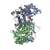 8gi7C 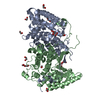 8giwC  8glbC 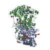 8gllC  8gmfC  8gmiC  8gmkC 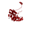 8s97C  8s9dC C: citing same article ( |
|---|---|
| Similar structure data |
-Links
 PDBj
PDBj

- Assembly
| Deposited unit | 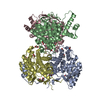
| ||||||||
|---|---|---|---|---|---|---|---|---|---|
| 1 | 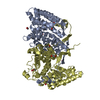
| ||||||||
| 2 | 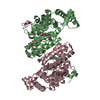
| ||||||||
| Unit cell |
|
-Components
| #1: Protein | Mass: 41019.605 Da / Num. of mol.: 4 Source method: isolated from a genetically manipulated source Source: (gene. exp.) Mycobacterium tuberculosis H37Rv (bacteria)Gene: citA, SAMEA2683035_02214 / Production host: References: UniProt: A0A045JB88, citrate synthase (unknown stereospecificity) #2: Chemical | ChemComp-EDO /   Mass: 62.068 Da / Num. of mol.: 12 / Source method: obtained synthetically / Formula: C2H6O2 Mass: 62.068 Da / Num. of mol.: 12 / Source method: obtained synthetically / Formula: C2H6O2#3: Chemical |   Mass: 106.120 Da / Num. of mol.: 3 / Source method: obtained synthetically / Formula: C4H10O3 Mass: 106.120 Da / Num. of mol.: 3 / Source method: obtained synthetically / Formula: C4H10O3#4: Chemical | ChemComp-AKR / 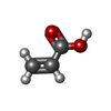  Mass: 72.063 Da / Num. of mol.: 4 / Source method: obtained synthetically / Formula: C3H4O2 Mass: 72.063 Da / Num. of mol.: 4 / Source method: obtained synthetically / Formula: C3H4O2#5: Water | ChemComp-HOH / |  Mass: 18.015 Da / Num. of mol.: 218 / Source method: isolated from a natural source / Formula: H2O Mass: 18.015 Da / Num. of mol.: 218 / Source method: isolated from a natural source / Formula: H2OHas ligand of interest | N | |
|---|
-Experimental details
-Experiment
| Experiment | Method: X-RAY DIFFRACTION / Number of used crystals: 1 |
|---|
- Sample preparation
| Crystal | Density Matthews: 2.76 Å3/Da / Density % sol: 55.39 % |
|---|---|
| Crystal grow | Temperature: 289 K / Method: vapor diffusion, hanging drop Details: 0.02 M magnesium chloride, 0.1 M HEPES pH 7.5, and 22 % w/v poly(acrylic acid sodium salt) 5100, 0.1 M barium chloride |
-Data collection
| Diffraction | Mean temperature: 100 K / Serial crystal experiment: N |
|---|---|
| Diffraction source | Source: SYNCHROTRON / Site: APS / Beamline: 21-ID-G / Wavelength: 0.9786 Å |
| Detector | Type: RAYONIX MX-300 / Detector: CCD / Date: Nov 29, 2012 |
| Radiation | Protocol: SINGLE WAVELENGTH / Monochromatic (M) / Laue (L): M / Scattering type: x-ray |
| Radiation wavelength | Wavelength: 0.9786 Å / Relative weight: 1 |
| Reflection | Resolution: 2.4→43.06 Å / Num. obs: 65711 / % possible obs: 94.26 % / Redundancy: 3.5 % / CC1/2: 0.982 / CC star: 0.995 / Rmerge(I) obs: 0.1686 / Rpim(I) all: 0.1063 / Rrim(I) all: 0.2 / Net I/σ(I): 6.93 |
| Reflection shell | Resolution: 2.4→2.486 Å / Redundancy: 3.8 % / Rmerge(I) obs: 1.057 / Num. unique obs: 6925 / CC1/2: 0.681 / CC star: 0.9 / Rpim(I) all: 0.6311 / Rrim(I) all: 1.233 / % possible all: 99.77 |
- Processing
| Software |
| ||||||||||||||||||||||||||||||||||||||||||||||||||||||||||||||||||||||||||||||||||||
|---|---|---|---|---|---|---|---|---|---|---|---|---|---|---|---|---|---|---|---|---|---|---|---|---|---|---|---|---|---|---|---|---|---|---|---|---|---|---|---|---|---|---|---|---|---|---|---|---|---|---|---|---|---|---|---|---|---|---|---|---|---|---|---|---|---|---|---|---|---|---|---|---|---|---|---|---|---|---|---|---|---|---|---|---|---|
| Refinement | Method to determine structure: MOLECULAR REPLACEMENT / Resolution: 2.4→43.06 Å / SU ML: 0.39 / Cross valid method: FREE R-VALUE / σ(F): 1.35 / Phase error: 31.95 / Stereochemistry target values: ML
| ||||||||||||||||||||||||||||||||||||||||||||||||||||||||||||||||||||||||||||||||||||
| Solvent computation | Shrinkage radii: 0.9 Å / VDW probe radii: 1.11 Å / Solvent model: FLAT BULK SOLVENT MODEL | ||||||||||||||||||||||||||||||||||||||||||||||||||||||||||||||||||||||||||||||||||||
| Refinement step | Cycle: LAST / Resolution: 2.4→43.06 Å
| ||||||||||||||||||||||||||||||||||||||||||||||||||||||||||||||||||||||||||||||||||||
| Refine LS restraints |
| ||||||||||||||||||||||||||||||||||||||||||||||||||||||||||||||||||||||||||||||||||||
| LS refinement shell |
|
