 Movie
Movie Controller


+ Open data

- Basic information
| Entry | Database: PDB / ID: 8gix | ||||||
|---|---|---|---|---|---|---|---|
| Title | Chaetomium thermophilum Hir3 | ||||||
 Components Components | Histone transcription regulator 3 homolog | ||||||
 Keywords Keywords | CHAPERONE / histone / complex / subunit | ||||||
| Function / homology |  Function and homology information Function and homology information | ||||||
| Biological species |  Thermochaetoides thermophila DSM 1495 (fungus) Thermochaetoides thermophila DSM 1495 (fungus) | ||||||
| Method | ELECTRON MICROSCOPY / single particle reconstruction / cryo EM / Resolution: 3.9 Å | ||||||
 Authors Authors | Szurgot, M.R. / van Eeuwen, T. / Kim, H.J. / Marmorstein, R. | ||||||
| Funding support |  United States, 1items United States, 1items
| ||||||
 Citation Citation | Journal: Mol Cell / Year: 2024 Title: Structure of the Hir histone chaperone complex. Authors: Hee Jong Kim / Mary R Szurgot / Trevor van Eeuwen / M Daniel Ricketts / Pratik Basnet / Athena L Zhang / Austin Vogt / Samah Sharmin / Craig D Kaplan / Benjamin A Garcia / Ronen Marmorstein / Kenji Murakami / Abstract: The evolutionarily conserved HIRA/Hir histone chaperone complex and ASF1a/Asf1 co-chaperone cooperate to deposit histone (H3/H4) tetramers on DNA for replication-independent chromatin assembly. The ...The evolutionarily conserved HIRA/Hir histone chaperone complex and ASF1a/Asf1 co-chaperone cooperate to deposit histone (H3/H4) tetramers on DNA for replication-independent chromatin assembly. The molecular architecture of the HIRA/Hir complex and its mode of histone deposition have remained unknown. Here, we report the cryo-EM structure of the S. cerevisiae Hir complex with Asf1/H3/H4 at 2.9-6.8 Å resolution. We find that the Hir complex forms an arc-shaped dimer with a Hir1/Hir2/Hir3/Hpc2 stoichiometry of 2/4/2/4. The core of the complex containing two Hir1/Hir2/Hir2 trimers and N-terminal segments of Hir3 forms a central cavity containing two copies of Hpc2, with one engaged by Asf1/H3/H4, in a suitable position to accommodate a histone (H3/H4) tetramer, while the C-terminal segments of Hir3 harbor nucleic acid binding activity to wrap DNA around the Hpc2-assisted histone tetramer. The structure suggests a model for how the Hir/Asf1 complex promotes the formation of histone tetramers for their subsequent deposition onto DNA. | ||||||
| History |
|
- Structure visualization
| Structure viewer | Molecule: MolmilJmol/JSmol |
|---|
- Downloads & links
-Download
| PDBx/mmCIF format | 8gix.cif.gz | 360.7 KB | Display | PDBx/mmCIF format |
|---|---|---|---|---|
| PDB format | pdb8gix.ent.gz | 263.8 KB | Display | PDB format |
| PDBx/mmJSON format | 8gix.json.gz | Tree view | PDBx/mmJSON format | |
| Others |  Other downloads Other downloads |
-Validation report
| Arichive directory | https://data.pdbj.org/pub/pdb/validation_reports/gi/8gixftp://data.pdbj.org/pub/pdb/validation_reports/gi/8gix | HTTPS FTP |
|---|
-Related structure data
| Related structure data | 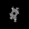 40078MC 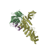 8ghaC 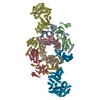 8ghlC 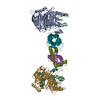 8ghmC 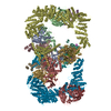 8ghnC M: map data used to model this data C: citing same article ( |
|---|---|
| Similar structure data |


-Links
 PDBj
PDBj


- Assembly
| Deposited unit | 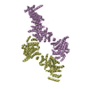
|
|---|---|
| 1 |
|
-Components
| #1: Protein | Mass: 250663.266 Da / Num. of mol.: 2 Source method: isolated from a genetically manipulated source Source: (gene. exp.) Thermochaetoides thermophila DSM 1495 (fungus)Strain: DSM 1495 / CBS 144.50 / IMI 039719 / Gene: CTHT_0035880 / Production host:  #2: Chemical | ChemComp-TCE / |   Mass: 250.186 Da / Num. of mol.: 1 / Source method: obtained synthetically / Formula: C9H15O6P Mass: 250.186 Da / Num. of mol.: 1 / Source method: obtained synthetically / Formula: C9H15O6PHas ligand of interest | N | |
|---|
-Experimental details
-Experiment
| Experiment | Method: ELECTRON MICROSCOPY |
|---|---|
| EM experiment | Aggregation state: PARTICLE / 3D reconstruction method: single particle reconstruction |
- Sample preparation
| Component | Name: Chaetomium thermophilum Hir3 / Type: COMPLEX / Entity ID: #1 / Source: RECOMBINANT |
|---|---|
| Source (natural) | Organism: Thermochaetoides thermophila (fungus) |
| Source (recombinant) | Organism: |
| Buffer solution | pH: 8 |
| Specimen | Embedding applied: NO / Shadowing applied: NO / Staining applied: NO / Vitrification applied: YES |
| Vitrification | Cryogen name: ETHANE |
- Electron microscopy imaging
| Experimental equipment |  Model: Titan Krios / Image courtesy: FEI Company |
|---|---|
| Microscopy | Model: FEI TITAN KRIOS |
| Electron gun | Electron source:  FIELD EMISSION GUN / Accelerating voltage: 300 kV / Illumination mode: FLOOD BEAM FIELD EMISSION GUN / Accelerating voltage: 300 kV / Illumination mode: FLOOD BEAM |
| Electron lens | Mode: BRIGHT FIELD / Nominal defocus max: 2600 nm / Nominal defocus min: 1000 nm |
| Image recording | Electron dose: 42.6 e/Å2 / Film or detector model: GATAN K3 (6k x 4k) |
- Processing
| EM software | Name: PHENIX / Version: 1.21_5207: / Category: model refinement |
|---|---|
| CTF correction | Type: PHASE FLIPPING AND AMPLITUDE CORRECTION |
| 3D reconstruction | Resolution: 3.9 Å / Resolution method: FSC 0.143 CUT-OFF / Num. of particles: 181856 / Symmetry type: POINT |
