 Movie
Movie Controller

[English] 日本語
 Yorodumi
- PDB-8gic: A1 Tei + Hpg: Adenylation domain 1 core construct from teicoplani... -
+ Open data

- Basic information
| Entry | Database: PDB / ID: 8gic | |||||||||
|---|---|---|---|---|---|---|---|---|---|---|
| Title | A1 Tei + Hpg: Adenylation domain 1 core construct from teicoplanin biosynthesis; 4-hydroxyphenylglycine bound | |||||||||
 Components Components |
| |||||||||
 Keywords Keywords | LIGASE / AMP-binding enzyme / NRPS / Adenylation domain | |||||||||
| Function / homology |  Function and homology information Function and homology informationsiderophore biosynthetic process / amino acid activation for nonribosomal peptide biosynthetic process / secondary metabolite biosynthetic process / lipid biosynthetic process / catalytic activity / phosphopantetheine binding / antibiotic biosynthetic process / cytosol Similarity search - Function | |||||||||
| Biological species |  Actinoplanes teichomyceticus (bacteria) Actinoplanes teichomyceticus (bacteria) | |||||||||
| Method |  X-RAY DIFFRACTION / SYNCHROTRON / MOLECULAR REPLACEMENT / Resolution: 1.64 Å X-RAY DIFFRACTION / SYNCHROTRON / MOLECULAR REPLACEMENT / Resolution: 1.64 Å | |||||||||
 Authors Authors | Hansen, M.H. / Cryle, M.J. | |||||||||
| Funding support |  Australia, 2items Australia, 2items
| |||||||||
 Citation Citation | Journal: Nat Commun / Year: 2023 Title: Resurrecting ancestral antibiotics: unveiling the origins of modern lipid II targeting glycopeptides. Authors: Hansen, M.H. / Adamek, M. / Iftime, D. / Petras, D. / Schuseil, F. / Grond, S. / Stegmann, E. / Cryle, M.J. / Ziemert, N. | |||||||||
| History |
|
- Structure visualization
| Structure viewer | Molecule: MolmilJmol/JSmol |
|---|
- Downloads & links
-Download
| PDBx/mmCIF format | 8gic.cif.gz | 433.9 KB | Display | PDBx/mmCIF format |
|---|---|---|---|---|
| PDB format | pdb8gic.ent.gz | 288.1 KB | Display | PDB format |
| PDBx/mmJSON format | 8gic.json.gz | Tree view | PDBx/mmJSON format | |
| Others |  Other downloads Other downloads |
-Validation report
| Arichive directory | https://data.pdbj.org/pub/pdb/validation_reports/gi/8gicftp://data.pdbj.org/pub/pdb/validation_reports/gi/8gic | HTTPS FTP |
|---|
-Related structure data
| Related structure data |  8gj4C  8gjpC  8gkmC  8glcC 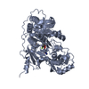 1amuS S: Starting model for refinement C: citing same article ( |
|---|---|
| Similar structure data |
-Links
 PDBj
PDBj


- Assembly
| Deposited unit | 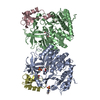
| ||||||||||||
|---|---|---|---|---|---|---|---|---|---|---|---|---|---|
| 1 | 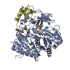
| ||||||||||||
| 2 | 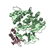
| ||||||||||||
| Unit cell |
|
-Components
| #1: Protein | Mass: 42084.703 Da / Num. of mol.: 2 Source method: isolated from a genetically manipulated source Source: (gene. exp.) Actinoplanes teichomyceticus (bacteria)Gene: tcp9 / Production host: #2: Protein | Mass: 7636.309 Da / Num. of mol.: 2 Source method: isolated from a genetically manipulated source Source: (gene. exp.) Actinoplanes teichomyceticus (bacteria)Gene: tcp13 / Production host: #3: Chemical |   Mass: 195.237 Da / Num. of mol.: 2 / Source method: isolated from a natural source / Formula: C6H13NO4S / Comment: pH buffer*YM Mass: 195.237 Da / Num. of mol.: 2 / Source method: isolated from a natural source / Formula: C6H13NO4S / Comment: pH buffer*YM#4: Chemical |   Type: L-peptide linking / Mass: 167.162 Da / Num. of mol.: 3 / Source method: isolated from a natural source / Formula: C8H9NO3 / Feature type: SUBJECT OF INVESTIGATION Type: L-peptide linking / Mass: 167.162 Da / Num. of mol.: 3 / Source method: isolated from a natural source / Formula: C8H9NO3 / Feature type: SUBJECT OF INVESTIGATION#5: Water | ChemComp-HOH / |  Mass: 18.015 Da / Num. of mol.: 771 / Source method: isolated from a natural source / Formula: H2O Mass: 18.015 Da / Num. of mol.: 771 / Source method: isolated from a natural source / Formula: H2OHas ligand of interest | Y | |
|---|
-Experimental details
-Experiment
| Experiment | Method: X-RAY DIFFRACTION / Number of used crystals: 1 |
|---|
- Sample preparation
| Crystal | Density Matthews: 2.32 Å3/Da / Density % sol: 46.99 % / Description: Cuboid |
|---|---|
| Crystal grow | Temperature: 293.15 K / Method: vapor diffusion, sitting drop / pH: 6 Details: 0.1 M MMT (L-Malic acid, MES and Tris in a ratio of 1:2:2), pH 6 and 25% w/v PEG 1500 |
-Data collection
| Diffraction | Mean temperature: 100 K / Serial crystal experiment: N |
|---|---|
| Diffraction source | Source: SYNCHROTRON / Site: Australian Synchrotron / Beamline: MX2 / Wavelength: 0.9537 Å |
| Detector | Type: DECTRIS EIGER X 16M / Detector: PIXEL / Date: Sep 25, 2018 |
| Radiation | Protocol: SINGLE WAVELENGTH / Monochromatic (M) / Laue (L): M / Scattering type: x-ray |
| Radiation wavelength | Wavelength: 0.9537 Å / Relative weight: 1 |
| Reflection | Resolution: 1.64→44.15 Å / Num. obs: 114836 / % possible obs: 99.98 % / Redundancy: 2 % / Biso Wilson estimate: 21.43 Å2 / CC1/2: 1 / Rmerge(I) obs: 0.02163 / Rpim(I) all: 0.02163 / Net I/σ(I): 14.32 |
| Reflection shell | Resolution: 1.64→1.699 Å / Rmerge(I) obs: 0.3679 / Mean I/σ(I) obs: 1.96 / Num. unique obs: 11325 / CC1/2: 0.767 / Rpim(I) all: 0.3679 / % possible all: 99.97 |
- Processing
| Software |
| |||||||||||||||||||||||||||||||||||||||||||||||||||||||||||||||||||||||||||||||||||||||||||||||||||||||||||||||||||||||||||||||||||||||||||||||||||||||||||||||||||||||||||||||||||||||||||||||||||||||||||||||||||||||||
|---|---|---|---|---|---|---|---|---|---|---|---|---|---|---|---|---|---|---|---|---|---|---|---|---|---|---|---|---|---|---|---|---|---|---|---|---|---|---|---|---|---|---|---|---|---|---|---|---|---|---|---|---|---|---|---|---|---|---|---|---|---|---|---|---|---|---|---|---|---|---|---|---|---|---|---|---|---|---|---|---|---|---|---|---|---|---|---|---|---|---|---|---|---|---|---|---|---|---|---|---|---|---|---|---|---|---|---|---|---|---|---|---|---|---|---|---|---|---|---|---|---|---|---|---|---|---|---|---|---|---|---|---|---|---|---|---|---|---|---|---|---|---|---|---|---|---|---|---|---|---|---|---|---|---|---|---|---|---|---|---|---|---|---|---|---|---|---|---|---|---|---|---|---|---|---|---|---|---|---|---|---|---|---|---|---|---|---|---|---|---|---|---|---|---|---|---|---|---|---|---|---|---|---|---|---|---|---|---|---|---|---|---|---|---|---|---|---|---|
| Refinement | Method to determine structure: MOLECULAR REPLACEMENT Starting model: 1AMU Resolution: 1.64→44.15 Å / SU ML: 0.1885 / Cross valid method: FREE R-VALUE / σ(F): 1.34 / Phase error: 19.7051 Stereochemistry target values: GeoStd + Monomer Library + CDL v1.2
| |||||||||||||||||||||||||||||||||||||||||||||||||||||||||||||||||||||||||||||||||||||||||||||||||||||||||||||||||||||||||||||||||||||||||||||||||||||||||||||||||||||||||||||||||||||||||||||||||||||||||||||||||||||||||
| Solvent computation | Shrinkage radii: 0.9 Å / VDW probe radii: 1.1 Å / Solvent model: FLAT BULK SOLVENT MODEL | |||||||||||||||||||||||||||||||||||||||||||||||||||||||||||||||||||||||||||||||||||||||||||||||||||||||||||||||||||||||||||||||||||||||||||||||||||||||||||||||||||||||||||||||||||||||||||||||||||||||||||||||||||||||||
| Displacement parameters | Biso mean: 27.12 Å2 | |||||||||||||||||||||||||||||||||||||||||||||||||||||||||||||||||||||||||||||||||||||||||||||||||||||||||||||||||||||||||||||||||||||||||||||||||||||||||||||||||||||||||||||||||||||||||||||||||||||||||||||||||||||||||
| Refinement step | Cycle: LAST / Resolution: 1.64→44.15 Å
| |||||||||||||||||||||||||||||||||||||||||||||||||||||||||||||||||||||||||||||||||||||||||||||||||||||||||||||||||||||||||||||||||||||||||||||||||||||||||||||||||||||||||||||||||||||||||||||||||||||||||||||||||||||||||
| Refine LS restraints |
| |||||||||||||||||||||||||||||||||||||||||||||||||||||||||||||||||||||||||||||||||||||||||||||||||||||||||||||||||||||||||||||||||||||||||||||||||||||||||||||||||||||||||||||||||||||||||||||||||||||||||||||||||||||||||
| LS refinement shell |
|
