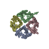
- PDB-8ghj: Crystal structure of human AQP2 T125M mutant -
+
Open data
ID or keywords:
Loading...
-
Basic information
Entry
Database: PDB / ID: 8ghj
Title
Crystal structure of human AQP2 T125M mutant
Components
Aquaporin-2
Keywords
MEMBRANE PROTEIN / Aquaporin / water channel
Function / homology
Function and homology information
cellular response to water deprivation / renal water transport / glycerol transmembrane transporter activity / water transmembrane transporter activity / Passive transport by Aquaporins / lumenal side of membrane / glycerol transmembrane transport / cellular response to mercury ion / water transport / water channel activity ...cellular response to water deprivation / renal water transport / glycerol transmembrane transporter activity / water transmembrane transporter activity / Passive transport by Aquaporins / lumenal side of membrane / glycerol transmembrane transport / cellular response to mercury ion / water transport / water channel activity / metanephric collecting duct development / transport vesicle membrane / renal water homeostasis / actin filament organization / cellular response to copper ion / recycling endosome / Vasopressin regulates renal water homeostasis via Aquaporins / protein homotetramerization / basolateral plasma membrane / apical plasma membrane / perinuclear region of cytoplasm / Golgi apparatus / extracellular exosome / membrane / plasma membrane Similarity search - Function
Aquaporin transporter / Major intrinsic protein, conserved site / MIP family signature. / Major intrinsic protein / Major intrinsic protein / Aquaporin-like Similarity search - Domain/homology
Aquaporin-2 / AQP-2 / ADH water channel / Aquaporin-CD / AQP-CD / Collecting duct water channel protein / WCH-CD ...AQP-2 / ADH water channel / Aquaporin-CD / AQP-CD / Collecting duct water channel protein / WCH-CD / Water channel protein for renal collecting duct
Mass: 25169.312 Da / Num. of mol.: 4 / Mutation: T125M Source method: isolated from a genetically manipulated source Source: (gene. exp.) Homo sapiens (human) / Gene: AQP2 / Production host: Komagataella pastoris (fungus) / References: UniProt: P41181
Mass: 112.411 Da / Num. of mol.: 2 / Source method: obtained synthetically / Formula: Cd
Has ligand of interest
N
Has protein modification
Y
-
Experimental details
-
Experiment
Experiment
Method: X-RAY DIFFRACTION / Number of used crystals: 1
-
Sample preparation
Crystal
Density Matthews: 3.26 Å3/Da / Density % sol: 62.29 %
Crystal grow
Temperature: 293 K / Method: vapor diffusion, hanging drop / pH: 8.5 Details: 20- 30% PEG400, Tris-HCl pH 8.5, 0.1M MgCl2, 0.1M NaCl, 0.025 M CdCl2 added to the drop only
-
Data collection
Diffraction
Mean temperature: 100 K / Serial crystal experiment: N
Diffraction source
Source: SYNCHROTRON / Site: MAX IV / Beamline: BioMAX / Wavelength: 0.977 Å
Detector
Type: DECTRIS EIGER X 16M / Detector: PIXEL / Date: Jan 24, 2019
Radiation
Protocol: SINGLE WAVELENGTH / Monochromatic (M) / Laue (L): M / Scattering type: x-ray
Radiation wavelength
Wavelength: 0.977 Å / Relative weight: 1
Reflection
Resolution: 3.9→50 Å / Num. obs: 22380 / % possible obs: 99.9 % / Redundancy: 21.3 % / Biso Wilson estimate: 215.48 Å2 / CC1/2: 0.99 / Net I/σ(I): 0.43
In the structure databanks used in Yorodumi, some data are registered as the other names, "COVID-19 virus" and "2019-nCoV". Here are the details of the virus and the list of structure data.
Jan 31, 2019. EMDB accession codes are about to change! (news from PDBe EMDB page)
EMDB accession codes are about to change! (news from PDBe EMDB page)
The allocation of 4 digits for EMDB accession codes will soon come to an end. Whilst these codes will remain in use, new EMDB accession codes will include an additional digit and will expand incrementally as the available range of codes is exhausted. The current 4-digit format prefixed with “EMD-” (i.e. EMD-XXXX) will advance to a 5-digit format (i.e. EMD-XXXXX), and so on. It is currently estimated that the 4-digit codes will be depleted around Spring 2019, at which point the 5-digit format will come into force.
The EM Navigator/Yorodumi systems omit the EMD- prefix.
Related info.:Q: What is EMD? / ID/Accession-code notation in Yorodumi/EM Navigator
Yorodumi is a browser for structure data from EMDB, PDB, SASBDB, etc.
This page is also the successor to EM Navigator detail page, and also detail information page/front-end page for Omokage search.
The word "yorodu" (or yorozu) is an old Japanese word meaning "ten thousand". "mi" (miru) is to see.
Related info.:EMDB / PDB / SASBDB / Comparison of 3 databanks / Yorodumi Search / Aug 31, 2016. New EM Navigator & Yorodumi / Yorodumi Papers / Jmol/JSmol / Function and homology information / Changes in new EM Navigator and Yorodumi
 Movie
Movie Controller


 Open data
 Basic information
 Components
 Keywords
 Function and homology information
 Homo sapiens (human)
 X-RAY DIFFRACTION /
  Authors
 Sweden, 2items
  Citation
 Structure visualization
 Downloads & links
 Other downloads
 PDBj
PDBj

 Assembly
 Komagataella pastoris (fungus) / References: UniProt: P41181
 Mass: 112.411 Da / Num. of mol.: 2 / Source method: obtained synthetically / Formula: Cd
 Sample preparation
 Processing