 Movie
Movie Controller

[English] 日本語
 Yorodumi
- PDB-8fm9: Nodavirus RNA replication proto-crown, detergent-solubliized C12 ... -
+ Open data

- Basic information
| Entry | Database: PDB / ID: 8fm9 | ||||||
|---|---|---|---|---|---|---|---|
| Title | Nodavirus RNA replication proto-crown, detergent-solubliized C12 multimer | ||||||
 Components Components | RNA-directed RNA polymerase | ||||||
 Keywords Keywords | VIRAL PROTEIN / Nodavirus RNA replication and RNA capping complex / Dodecamer ring / Outer mitochondrial membrane protein complex | ||||||
| Function / homology | Nodavirus methyltransferase domain / Nodavirus Vmethyltransferase / host cell mitochondrial outer membrane / RNA-directed RNA polymerase / RNA-directed RNA polymerase activity / DNA/RNA polymerase superfamily / RNA-directed RNA polymerase Function and homology information Function and homology information | ||||||
| Biological species |  Flock House virus Flock House virus | ||||||
| Method | ELECTRON MICROSCOPY / single particle reconstruction / cryo EM / Resolution: 3.2 Å | ||||||
 Authors Authors | Zhan, H. / Unchwaniwala, N. / Rebolledo Viveros, A. / Pennington, J. / Horswill, M. / Broadberry, R. / Myers, J. / den Boon, J. / Grant, T. / Ahlquist, P. | ||||||
| Funding support |  United States, 1items United States, 1items
| ||||||
 Citation Citation | Journal: Proc Natl Acad Sci U S A / Year: 2023 Title: Nodavirus RNA replication crown architecture reveals proto-crown precursor and viral protein A conformational switching. Authors: Hong Zhan / Nuruddin Unchwaniwala / Andrea Rebolledo-Viveros / Janice Pennington / Mark Horswill / Roma Broadberry / Jonathan Myers / Johan A den Boon / Timothy Grant / Paul Ahlquist / Abstract: Positive-strand RNA viruses replicate their genomes in virus-induced membrane vesicles, and the resulting RNA replication complexes are a major target for virus control. Nodavirus studies first ...Positive-strand RNA viruses replicate their genomes in virus-induced membrane vesicles, and the resulting RNA replication complexes are a major target for virus control. Nodavirus studies first revealed viral RNA replication proteins forming a 12-fold symmetric "crown" at the vesicle opening to the cytosol, an arrangement recently confirmed to extend to distantly related alphaviruses. Using cryoelectron microscopy (cryo-EM), we show that mature nodavirus crowns comprise two stacked 12-mer rings of multidomain viral RNA replication protein A. Each ring contains an ~19 nm circle of C-proximal polymerase domains, differentiated by strikingly diverged positions of N-proximal RNA capping/membrane binding domains. The lower ring is a "proto-crown" precursor that assembles prior to RNA template recruitment, RNA synthesis, and replication vesicle formation. In this proto-crown, the N-proximal segments interact to form a toroidal central floor, whose 3.1 Å resolution structure reveals many mechanistic details of the RNA capping/membrane binding domains. In the upper ring, cryo-EM fitting indicates that the N-proximal domains extend radially outside the polymerases, forming separated, membrane-binding "legs." The polymerase and N-proximal domains are connected by a long linker accommodating the conformational switch between the two rings and possibly also polymerase movements associated with RNA synthesis and nonsymmetric electron density in the lower center of mature crowns. The results reveal remarkable viral protein multifunctionality, conformational flexibility, and evolutionary plasticity and insights into (+)RNA virus replication and control. | ||||||
| History |
|
- Structure visualization
| Structure viewer | Molecule: MolmilJmol/JSmol |
|---|
- Downloads & links
-Download
| PDBx/mmCIF format | 8fm9.cif.gz | 1.9 MB | Display | PDBx/mmCIF format |
|---|---|---|---|---|
| PDB format | pdb8fm9.ent.gz | 1.4 MB | Display | PDB format |
| PDBx/mmJSON format | 8fm9.json.gz | Tree view | PDBx/mmJSON format | |
| Others |  Other downloads Other downloads |
-Validation report
| Arichive directory | https://data.pdbj.org/pub/pdb/validation_reports/fm/8fm9ftp://data.pdbj.org/pub/pdb/validation_reports/fm/8fm9 | HTTPS FTP |
|---|
-Related structure data
| Related structure data |  29289MC 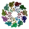 8fmaC 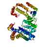 8fmbC M: map data used to model this data C: citing same article ( |
|---|---|
| Similar structure data |


-Links
 PDBj
PDBj- Assembly
| Deposited unit | 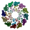
|
|---|---|
| 1 |
|
-Components
| #1: Protein | Mass: 113996.586 Da / Num. of mol.: 24 Source method: isolated from a genetically manipulated source Source: (gene. exp.) Flock House virus / Cell line (production host): Sf9 / Production host:   Spodoptera frugiperda (fall armyworm) / References: UniProt: Q66929, RNA-directed RNA polymerase Spodoptera frugiperda (fall armyworm) / References: UniProt: Q66929, RNA-directed RNA polymerase |
|---|
-Experimental details
-Experiment
| Experiment | Method: ELECTRON MICROSCOPY |
|---|---|
| EM experiment | Aggregation state: PARTICLE / 3D reconstruction method: single particle reconstruction |
- Sample preparation
| Component | Name: Nodavirus RNA replication proto-crown, detergent-solubliized C12 multimer Type: COMPLEX / Entity ID: all / Source: RECOMBINANT |
|---|---|
| Molecular weight | Experimental value: NO |
| Source (natural) | Organism: Flock House virus |
| Source (recombinant) | Organism: Spodoptera frugiperda (fall armyworm) |
| Buffer solution | pH: 7.4 |
| Specimen | Embedding applied: NO / Shadowing applied: NO / Staining applied: NO / Vitrification applied: YES |
| Specimen support | Grid material: COPPER / Grid mesh size: 300 divisions/in. / Grid type: Quantifoil R1.2/1.3 |
| Vitrification | Instrument: FEI VITROBOT MARK IV / Cryogen name: ETHANE / Humidity: 100 % / Chamber temperature: 277 K |
- Electron microscopy imaging
| Experimental equipment |  Model: Titan Krios / Image courtesy: FEI Company |
|---|---|
| Microscopy | Model: FEI TITAN KRIOS |
| Electron gun | Electron source:  FIELD EMISSION GUN / Accelerating voltage: 300 kV / Illumination mode: FLOOD BEAM FIELD EMISSION GUN / Accelerating voltage: 300 kV / Illumination mode: FLOOD BEAM |
| Electron lens | Mode: BRIGHT FIELD / Nominal defocus max: 2500 nm / Nominal defocus min: 500 nm / Cs: 2.7 mm / C2 aperture diameter: 100 µm |
| Image recording | Electron dose: 100 e/Å2 / Detector mode: INTEGRATING / Film or detector model: FEI FALCON III (4k x 4k) |
| Image scans | Width: 4096 / Height: 4096 |
- Processing
| Software | Name: PHENIX / Version: 1.20.1_4487: / Classification: refinement | ||||||||||||||||||||||||||||||||||||||||||||||||
|---|---|---|---|---|---|---|---|---|---|---|---|---|---|---|---|---|---|---|---|---|---|---|---|---|---|---|---|---|---|---|---|---|---|---|---|---|---|---|---|---|---|---|---|---|---|---|---|---|---|
| EM software |
| ||||||||||||||||||||||||||||||||||||||||||||||||
| CTF correction | Type: PHASE FLIPPING AND AMPLITUDE CORRECTION | ||||||||||||||||||||||||||||||||||||||||||||||||
| Symmetry | Point symmetry: C12 (12 fold cyclic) | ||||||||||||||||||||||||||||||||||||||||||||||||
| 3D reconstruction | Resolution: 3.2 Å / Resolution method: FSC 0.143 CUT-OFF / Num. of particles: 11093 / Symmetry type: POINT | ||||||||||||||||||||||||||||||||||||||||||||||||
| Atomic model building | Protocol: FLEXIBLE FIT Details: The final model includes 24 chains representing 12 protein A polypeptides. Chains A-L represent the N-terminal floor segment built into the high-resolution EM density. Chains M-X represent a ...Details: The final model includes 24 chains representing 12 protein A polypeptides. Chains A-L represent the N-terminal floor segment built into the high-resolution EM density. Chains M-X represent a mainchain trace of the polymerase segment rigid body fit into the map and are included to give a more complete picture of the molecule. The polymerase mainchain trace was refined from an Alphafold prediction into a map obtained by symmetry expansion with subtraction. The polymerase map is uploaded as a separate entry. The two segments have separate chain IDs as it is unclear from the density which polymerase connects to which floor segment. | ||||||||||||||||||||||||||||||||||||||||||||||||
| Refine LS restraints |
|
