 Movie
Movie Controller

[English] 日本語
 Yorodumi
- PDB-8fhw: Cryo-EM structure of Cryptococcus neoformans trehalose-6-phosphat... -
+ Open data

- Basic information
| Entry | Database: PDB / ID: 8fhw | ||||||
|---|---|---|---|---|---|---|---|
| Title | Cryo-EM structure of Cryptococcus neoformans trehalose-6-phosphate synthase homotetramer in complex with uridine diphosphate and glucose-6-phosphate | ||||||
 Components Components | Alpha,alpha-trehalose-phosphate synthase (UDP-forming) | ||||||
 Keywords Keywords | TRANSFERASE / Glycosyltransferase / Complex | ||||||
| Function / homology |  Function and homology information Function and homology informationalpha,alpha-trehalose-phosphate synthase complex (UDP-forming) / alpha,alpha-trehalose-phosphate synthase (UDP-forming) / trehalose-phosphatase activity / alpha,alpha-trehalose-phosphate synthase (UDP-forming) activity / trehalose biosynthetic process / cellular response to heat / cytosol Similarity search - Function | ||||||
| Biological species |  Cryptococcus neoformans var. grubii H99 (fungus) Cryptococcus neoformans var. grubii H99 (fungus) | ||||||
| Method | ELECTRON MICROSCOPY / single particle reconstruction / cryo EM / Resolution: 3.2 Å | ||||||
 Authors Authors | Washington, E.J. / Brennan, R.G. | ||||||
| Funding support |  United States, 1items United States, 1items
| ||||||
 Citation Citation | Journal: bioRxiv / Year: 2024 Title: Structures of trehalose-6-phosphate synthase, Tps1, from the fungal pathogen : a target for novel antifungals. Authors: Erica J Washington / Ye Zhou / Allen L Hsu / Matthew Petrovich / Jennifer L Tenor / Dena L Toffaletti / Ziqiang Guan / John R Perfect / Mario J Borgnia / Alberto Bartesaghi / Richard G Brennan Abstract: Invasive fungal diseases are a major threat to human health, resulting in more than 1.5 million annual deaths worldwide. The arsenal of antifungal therapeutics remains limited and is in dire need of ...Invasive fungal diseases are a major threat to human health, resulting in more than 1.5 million annual deaths worldwide. The arsenal of antifungal therapeutics remains limited and is in dire need of novel drugs that target additional biosynthetic pathways that are absent from humans. One such pathway involves the biosynthesis of trehalose. Trehalose is a disaccharide that is required for pathogenic fungi to survive in their human hosts. In the first step of trehalose biosynthesis, trehalose-6-phosphate synthase (Tps1) converts UDP-glucose and glucose-6-phosphate to trehalose-6-phosphate. Here, we report the structures of full-length Tps1 (CnTps1) in unliganded form and in complex with uridine diphosphate and glucose-6-phosphate. Comparison of these two structures reveals significant movement towards the catalytic pocket by the N-terminus upon ligand binding and identifies residues required for substrate-binding, as well as residues that stabilize the tetramer. Intriguingly, an intrinsically disordered domain (IDD), which is conserved amongst Cryptococcal species and closely related Basidiomycetes, extends from each subunit of the tetramer into the "solvent" but is not visible in density maps. We determined that the IDD is not required for Tps1-dependent thermotolerance and osmotic stress survival. Studies with UDP-galactose highlight the exquisite substrate specificity of CnTps1. , these studies expand our knowledge of trehalose biosynthesis in and highlight the potential of developing antifungal therapeutics that disrupt the synthesis of this disaccharide or the formation of a functional tetramer and the use of cryo-EM in the structural characterization of CnTps1-ligand/drug complexes. SIGNIFICANCE STATEMENT: Fungal infections are responsible for over a million deaths worldwide each year. Biosynthesis of a disaccharide, trehalose, is required for multiple pathogenic fungi to ...SIGNIFICANCE STATEMENT: Fungal infections are responsible for over a million deaths worldwide each year. Biosynthesis of a disaccharide, trehalose, is required for multiple pathogenic fungi to transition from the environment to the human host. Enzymes in the trehalose biosynthesis pathway are absent in humans and, therefore, are potentially significant targets for novel antifungal therapeutics. One enzyme in the trehalose biosynthesis is trehalose-6-phosphate synthase (Tps1). Here, we describe the cryo-electron microscopy structures of the CnTps1 homo-tetramer in the unliganded form and in complex with a substrate and a product. These structures and subsequent biochemical analysis reveal key details of substrate-binding residues and substrate specificity. These structures should facilitate structure-guided design of inhibitors against CnTps1. | ||||||
| History |
|
- Structure visualization
| Structure viewer | Molecule: MolmilJmol/JSmol |
|---|
- Downloads & links
-Download
| PDBx/mmCIF format | 8fhw.cif.gz | 563.8 KB | Display | PDBx/mmCIF format |
|---|---|---|---|---|
| PDB format | pdb8fhw.ent.gz | 461.2 KB | Display | PDB format |
| PDBx/mmJSON format | 8fhw.json.gz | Tree view | PDBx/mmJSON format | |
| Others |  Other downloads Other downloads |
-Validation report
| Arichive directory | https://data.pdbj.org/pub/pdb/validation_reports/fh/8fhwftp://data.pdbj.org/pub/pdb/validation_reports/fh/8fhw | HTTPS FTP |
|---|
-Related structure data
| Related structure data |  29172MC M: map data used to model this data C: citing same article ( |
|---|---|
| Similar structure data |
-Links
 PDBj
PDBj


- Assembly
| Deposited unit | 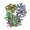
|
|---|---|
| 1 |
|
-Components
| #1: Protein | Mass: 76815.500 Da / Num. of mol.: 4 Source method: isolated from a genetically manipulated source Details: UDP G6P Source: (gene. exp.) Cryptococcus neoformans var. grubii H99 (fungus)Gene: CNAG_05292 / Plasmid: pMCSG7 / Production host:  #2: Chemical | ChemComp-UDP /   Type: RNA linking / Mass: 404.161 Da / Num. of mol.: 4 / Source method: obtained synthetically / Formula: C9H14N2O12P2 / Feature type: SUBJECT OF INVESTIGATION / Comment: UDP*YM Type: RNA linking / Mass: 404.161 Da / Num. of mol.: 4 / Source method: obtained synthetically / Formula: C9H14N2O12P2 / Feature type: SUBJECT OF INVESTIGATION / Comment: UDP*YM#3: Sugar | ChemComp-G6P /   Type: D-saccharide, alpha linking / Mass: 260.136 Da / Num. of mol.: 4 / Source method: obtained synthetically / Formula: C6H13O9P / Feature type: SUBJECT OF INVESTIGATION Type: D-saccharide, alpha linking / Mass: 260.136 Da / Num. of mol.: 4 / Source method: obtained synthetically / Formula: C6H13O9P / Feature type: SUBJECT OF INVESTIGATIONHas ligand of interest | Y | |
|---|
-Experimental details
-Experiment
| Experiment | Method: ELECTRON MICROSCOPY |
|---|---|
| EM experiment | Aggregation state: PARTICLE / 3D reconstruction method: single particle reconstruction |
- Sample preparation
| Component | Name: Cryo-EM structure of Cryptococcus neoformans trehalose-6-phosphate synthase homotetramer in complex with uridine diphosphate and glucose-6-phosphate Type: COMPLEX / Entity ID: #1 / Source: RECOMBINANT |
|---|---|
| Molecular weight | Value: 0.307 MDa / Experimental value: YES |
| Source (natural) | Organism: Cryptococcus neoformans var. grubii H99 (fungus) |
| Source (recombinant) | Organism: |
| Buffer solution | pH: 7.5 |
| Specimen | Conc.: 0.75 mg/ml / Embedding applied: NO / Shadowing applied: NO / Staining applied: NO / Vitrification applied: YES |
| Vitrification | Cryogen name: ETHANE |
- Electron microscopy imaging
| Experimental equipment |  Model: Titan Krios / Image courtesy: FEI Company |
|---|---|
| Microscopy | Model: FEI TITAN KRIOS |
| Electron gun | Electron source:  FIELD EMISSION GUN / Accelerating voltage: 300 kV / Illumination mode: OTHER FIELD EMISSION GUN / Accelerating voltage: 300 kV / Illumination mode: OTHER |
| Electron lens | Mode: BRIGHT FIELD / Nominal defocus max: 2500 nm / Nominal defocus min: 800 nm |
| Image recording | Electron dose: 60 e/Å2 / Film or detector model: GATAN K3 BIOQUANTUM (6k x 4k) |
- Processing
| CTF correction | Type: PHASE FLIPPING AND AMPLITUDE CORRECTION | ||||||||||||||||||||||||
|---|---|---|---|---|---|---|---|---|---|---|---|---|---|---|---|---|---|---|---|---|---|---|---|---|---|
| 3D reconstruction | Resolution: 3.2 Å / Resolution method: FSC 0.143 CUT-OFF / Num. of particles: 854608 / Symmetry type: POINT | ||||||||||||||||||||||||
| Refinement | Cross valid method: NONE Stereochemistry target values: GeoStd + Monomer Library + CDL v1.2 | ||||||||||||||||||||||||
| Displacement parameters | Biso mean: 48.64 Å2 | ||||||||||||||||||||||||
| Refine LS restraints |
|
