 Movie
Movie Controller


+ Open data

- Basic information
| Entry | Database: PDB / ID: 8f7s | |||||||||||||||||||||||||||||||||||||||||||||||||||||||||
|---|---|---|---|---|---|---|---|---|---|---|---|---|---|---|---|---|---|---|---|---|---|---|---|---|---|---|---|---|---|---|---|---|---|---|---|---|---|---|---|---|---|---|---|---|---|---|---|---|---|---|---|---|---|---|---|---|---|---|
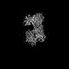
| Title | Gi bound delta-opioid receptor in complex with deltorphin | |||||||||||||||||||||||||||||||||||||||||||||||||||||||||
 Components Components |
| |||||||||||||||||||||||||||||||||||||||||||||||||||||||||
 Keywords Keywords | SIGNALING PROTEIN / delta opioid receptor / G protein coupled receptor / deltorphin | |||||||||||||||||||||||||||||||||||||||||||||||||||||||||
| Function / homology |  Function and homology information Function and homology informationopioid peptide activity / G protein-coupled enkephalin receptor activity / spine apparatus / G protein-coupled opioid receptor activity / G protein-coupled opioid receptor signaling pathway / receptor serine/threonine kinase binding / cellular response to toxic substance / G-protein activation / Activation of the phototransduction cascade / Glucagon-type ligand receptors ...opioid peptide activity / G protein-coupled enkephalin receptor activity / spine apparatus / G protein-coupled opioid receptor activity / G protein-coupled opioid receptor signaling pathway / receptor serine/threonine kinase binding / cellular response to toxic substance / G-protein activation / Activation of the phototransduction cascade / Glucagon-type ligand receptors / Thromboxane signalling through TP receptor / Sensory perception of sweet, bitter, and umami (glutamate) taste / G beta:gamma signalling through PI3Kgamma / G beta:gamma signalling through CDC42 / Cooperation of PDCL (PhLP1) and TRiC/CCT in G-protein beta folding / Activation of G protein gated Potassium channels / Inhibition of voltage gated Ca2+ channels via Gbeta/gamma subunits / Ca2+ pathway / G alpha (z) signalling events / High laminar flow shear stress activates signaling by PIEZO1 and PECAM1:CDH5:KDR in endothelial cells / Glucagon-like Peptide-1 (GLP1) regulates insulin secretion / Vasopressin regulates renal water homeostasis via Aquaporins / Adrenaline,noradrenaline inhibits insulin secretion / ADP signalling through P2Y purinoceptor 12 / G alpha (q) signalling events / G alpha (i) signalling events / Thrombin signalling through proteinase activated receptors (PARs) / Activation of G protein gated Potassium channels / G-protein activation / G beta:gamma signalling through PI3Kgamma / Prostacyclin signalling through prostacyclin receptor / G beta:gamma signalling through PLC beta / ADP signalling through P2Y purinoceptor 1 / Thromboxane signalling through TP receptor / Presynaptic function of Kainate receptors / G beta:gamma signalling through CDC42 / Inhibition of voltage gated Ca2+ channels via Gbeta/gamma subunits / neuropeptide binding / G alpha (12/13) signalling events / Glucagon-type ligand receptors / G beta:gamma signalling through BTK / ADP signalling through P2Y purinoceptor 12 / Adrenaline,noradrenaline inhibits insulin secretion / Cooperation of PDCL (PhLP1) and TRiC/CCT in G-protein beta folding / Ca2+ pathway / G alpha (z) signalling events / Thrombin signalling through proteinase activated receptors (PARs) / Extra-nuclear estrogen signaling / G alpha (s) signalling events / G alpha (q) signalling events / photoreceptor outer segment membrane / spectrin binding / Glucagon-like Peptide-1 (GLP1) regulates insulin secretion / G alpha (i) signalling events / High laminar flow shear stress activates signaling by PIEZO1 and PECAM1:CDH5:KDR in endothelial cells / Vasopressin regulates renal water homeostasis via Aquaporins / eating behavior / alkylglycerophosphoethanolamine phosphodiesterase activity / regulation of calcium ion transport / photoreceptor outer segment / G protein-coupled receptor signaling pathway, coupled to cyclic nucleotide second messenger / neuronal dense core vesicle / negative regulation of protein-containing complex assembly / cardiac muscle cell apoptotic process / adenylate cyclase inhibitor activity / photoreceptor inner segment / positive regulation of protein localization to cell cortex / T cell migration / positive regulation of relaxation of smooth muscle / Adenylate cyclase inhibitory pathway / D2 dopamine receptor binding / adenylate cyclase-inhibiting serotonin receptor signaling pathway / G protein-coupled serotonin receptor binding / dendrite membrane / axon terminus / cellular response to forskolin / Peptide ligand-binding receptors / regulation of mitotic spindle organization / chemokine-mediated signaling pathway / response to nicotine / regulation of mitochondrial membrane potential / adult locomotory behavior / defense response / Regulation of insulin secretion / neuropeptide signaling pathway / response to prostaglandin E / positive regulation of cholesterol biosynthetic process / negative regulation of insulin secretion / postsynaptic density membrane / G protein-coupled receptor binding / cellular response to growth factor stimulus / response to peptide hormone / centriolar satellite / G-protein beta/gamma-subunit complex binding / adenylate cyclase-modulating G protein-coupled receptor signaling pathway / adenylate cyclase-inhibiting G protein-coupled receptor signaling pathway / ADP signalling through P2Y purinoceptor 12 / GDP binding / Adrenaline,noradrenaline inhibits insulin secretion / G alpha (z) signalling events Similarity search - Function | |||||||||||||||||||||||||||||||||||||||||||||||||||||||||
| Biological species |  Homo sapiens (human) Homo sapiens (human) Phyllomedusa (leaf frogs) | |||||||||||||||||||||||||||||||||||||||||||||||||||||||||
| Method | ELECTRON MICROSCOPY / single particle reconstruction / cryo EM / Resolution: 3 Å | |||||||||||||||||||||||||||||||||||||||||||||||||||||||||
 Authors Authors | Wang, Y. / Zhuang, Y. / DiBerto, J.F. / Zhou, X.E. / Schmitz, G.P. / Yuan, Q. / Jain, M.K. / Liu, W. / Melcher, K. / Jiang, Y. ...Wang, Y. / Zhuang, Y. / DiBerto, J.F. / Zhou, X.E. / Schmitz, G.P. / Yuan, Q. / Jain, M.K. / Liu, W. / Melcher, K. / Jiang, Y. / Roth, B.L. / Xu, H.E. | |||||||||||||||||||||||||||||||||||||||||||||||||||||||||
| Funding support |  China, 3items China, 3items
| |||||||||||||||||||||||||||||||||||||||||||||||||||||||||
 Citation Citation | Journal: Cell / Year: 2023 Title: Structures of the entire human opioid receptor family. Authors: Yue Wang / Youwen Zhuang / Jeffrey F DiBerto / X Edward Zhou / Gavin P Schmitz / Qingning Yuan / Manish K Jain / Weiyi Liu / Karsten Melcher / Yi Jiang / Bryan L Roth / H Eric Xu /  Abstract: Opioids are effective analgesics, but their use is beset by serious side effects, including addiction and respiratory depression, which contribute to the ongoing opioid crisis. The human opioid ...Opioids are effective analgesics, but their use is beset by serious side effects, including addiction and respiratory depression, which contribute to the ongoing opioid crisis. The human opioid system contains four opioid receptors (μOR, δOR, κOR, and NOPR) and a set of related endogenous opioid peptides (EOPs), which show distinct selectivity toward their respective opioid receptors (ORs). Despite being key to the development of safer analgesics, the mechanisms of molecular recognition and selectivity of EOPs to ORs remain unclear. Here, we systematically characterize the binding of EOPs to ORs and present five structures of EOP-OR-G complexes, including β-endorphin- and endomorphin-bound μOR, deltorphin-bound δOR, dynorphin-bound κOR, and nociceptin-bound NOPR. These structures, supported by biochemical results, uncover the specific recognition and selectivity of opioid peptides and the conserved mechanism of opioid receptor activation. These results provide a structural framework to facilitate rational design of safer opioid drugs for pain relief. | |||||||||||||||||||||||||||||||||||||||||||||||||||||||||
| History |
|
- Structure visualization
| Structure viewer | Molecule: MolmilJmol/JSmol |
|---|
- Downloads & links
-Download
| PDBx/mmCIF format | 8f7s.cif.gz | 244.9 KB | Display | PDBx/mmCIF format |
|---|---|---|---|---|
| PDB format | pdb8f7s.ent.gz | 187.6 KB | Display | PDB format |
| PDBx/mmJSON format | 8f7s.json.gz | Tree view | PDBx/mmJSON format | |
| Others |  Other downloads Other downloads |
-Validation report
| Arichive directory | https://data.pdbj.org/pub/pdb/validation_reports/f7/8f7sftp://data.pdbj.org/pub/pdb/validation_reports/f7/8f7s | HTTPS FTP |
|---|
-Related structure data
| Related structure data |  28909MC 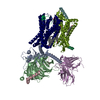 8f7qC 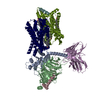 8f7rC 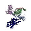 8f7wC 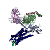 8f7xC M: map data used to model this data C: citing same article ( |
|---|---|
| Similar structure data |


-Links
 PDBj
PDBj


- Assembly
| Deposited unit | 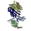
|
|---|---|
| 1 |
|
-Components
-Guanine nucleotide-binding protein ... , 3 types, 4 molecules AFBC
| #3: Protein | Mass: 40445.059 Da / Num. of mol.: 2 Source method: isolated from a genetically manipulated source Source: (gene. exp.) Homo sapiens (human) / Gene: GNAI1 / Production host:   Spodoptera frugiperda (fall armyworm) / References: UniProt: P63096 Spodoptera frugiperda (fall armyworm) / References: UniProt: P63096#4: Protein | | Mass: 39020.664 Da / Num. of mol.: 1 Source method: isolated from a genetically manipulated source Source: (gene. exp.) Spodoptera frugiperda (fall armyworm) / References: UniProt: P54311#5: Protein | | Mass: 7563.750 Da / Num. of mol.: 1 Source method: isolated from a genetically manipulated source Source: (gene. exp.) Spodoptera frugiperda (fall armyworm) / References: UniProt: P63212 |
|---|
-Protein / Protein/peptide , 2 types, 4 molecules RDPQ
| #1: Protein | Mass: 42651.020 Da / Num. of mol.: 2 Source method: isolated from a genetically manipulated source Source: (gene. exp.) Homo sapiens (human) / Gene: OPRD1, OPRD / Production host: Spodoptera frugiperda (fall armyworm) / References: UniProt: P41143#2: Protein/peptide | Mass: 781.876 Da / Num. of mol.: 2 / Fragment: UNP residues 107-113 / Source method: obtained synthetically / Source: (synth.) Phyllomedusa (leaf frogs) / References: UniProt: P21850 |
|---|
-Non-polymers , 2 types, 17 molecules 

| #6: Chemical | ChemComp-CLR /  Mass: 386.654 Da / Num. of mol.: 5 / Source method: obtained synthetically / Formula: C27H46O Mass: 386.654 Da / Num. of mol.: 5 / Source method: obtained synthetically / Formula: C27H46O#7: Chemical | ChemComp-PLM /  Mass: 256.424 Da / Num. of mol.: 12 / Source method: obtained synthetically / Formula: C16H32O2 Mass: 256.424 Da / Num. of mol.: 12 / Source method: obtained synthetically / Formula: C16H32O2 |
|---|
-Details
| Has ligand of interest | N |
|---|---|
| Has protein modification | Y |
-Experimental details
-Experiment
| Experiment | Method: ELECTRON MICROSCOPY |
|---|---|
| EM experiment | Aggregation state: PARTICLE / 3D reconstruction method: single particle reconstruction |
- Sample preparation
| Component | Name: Gi bound mu-opioid receptor in complex with beta-endorphin Type: COMPLEX / Entity ID: #1-#5 / Source: MULTIPLE SOURCES |
|---|---|
| Source (natural) | Organism: Homo sapiens (human) |
| Source (recombinant) | Organism: Spodoptera frugiperda (fall armyworm) |
| Buffer solution | pH: 7.2 |
| Specimen | Embedding applied: NO / Shadowing applied: NO / Staining applied: NO / Vitrification applied: YES |
| Vitrification | Cryogen name: ETHANE |
- Electron microscopy imaging
| Experimental equipment |  Model: Titan Krios / Image courtesy: FEI Company |
|---|---|
| Microscopy | Model: FEI TITAN KRIOS |
| Electron gun | Electron source:  FIELD EMISSION GUN / Accelerating voltage: 300 kV / Illumination mode: FLOOD BEAM FIELD EMISSION GUN / Accelerating voltage: 300 kV / Illumination mode: FLOOD BEAM |
| Electron lens | Mode: BRIGHT FIELD / Nominal defocus max: 1800 nm / Nominal defocus min: 800 nm |
| Image recording | Electron dose: 23.3 e/Å2 / Film or detector model: GATAN K3 (6k x 4k) |
- Processing
| Software | Name: PHENIX / Version: 1.13_2998: / Classification: refinement |
|---|---|
| EM software | Name: PHENIX / Category: model refinement |
| CTF correction | Type: NONE |
| 3D reconstruction | Resolution: 3 Å / Resolution method: FSC 0.143 CUT-OFF / Num. of particles: 650970 / Symmetry type: POINT |
