 Movie
Movie Controller

[English] 日本語
 Yorodumi
- PDB-8f67: Crystal structure of the refolded Penicillin Binding Protein 5 (P... -
+ Open data

- Basic information
| Entry | Database: PDB / ID: 8f67 | ||||||
|---|---|---|---|---|---|---|---|
| Title | Crystal structure of the refolded Penicillin Binding Protein 5 (PBP5) of Enterococcus faecium | ||||||
 Components Components | Pbp5 | ||||||
 Keywords Keywords | ANTIBIOTIC / Penicillin binding / antibiotic resistance / antibiotics | ||||||
| Function / homology |  Function and homology information Function and homology informationpeptidoglycan L,D-transpeptidase activity / penicillin binding / cell wall organization / response to antibiotic / plasma membrane Similarity search - Function | ||||||
| Biological species |  Enterococcus faecium (bacteria) Enterococcus faecium (bacteria) | ||||||
| Method |  X-RAY DIFFRACTION / SYNCHROTRON / MOLECULAR REPLACEMENT / Resolution: 3.59 Å X-RAY DIFFRACTION / SYNCHROTRON / MOLECULAR REPLACEMENT / Resolution: 3.59 Å | ||||||
 Authors Authors | D'Andrea, E.D. / Schoenle, M.V. / Choy, M.S. / Peti, W. / Page, R. | ||||||
| Funding support |  United States, 1items United States, 1items
| ||||||
 Citation Citation | Journal: Nat Commun / Year: 2023 Title: The Molecular Basis for Resistance of E. faecium PBP5 to beta-lactam Antibiotics Authors: Hunashal, Y. / Kumar, G.S. / Choy, M.S. / Da Silva Santiago, A. / D'Andrea, E.D. / Schoenle, M.V. / Arthur, M. / Rice, L.B. / Page, R. / Peti, W. | ||||||
| History |
|
- Structure visualization
| Structure viewer | Molecule: MolmilJmol/JSmol |
|---|
- Downloads & links
-Download
| PDBx/mmCIF format | 8f67.cif.gz | 373.5 KB | Display | PDBx/mmCIF format |
|---|---|---|---|---|
| PDB format | pdb8f67.ent.gz | 303.4 KB | Display | PDB format |
| PDBx/mmJSON format | 8f67.json.gz | Tree view | PDBx/mmJSON format | |
| Others |  Other downloads Other downloads |
-Validation report
| Arichive directory | https://data.pdbj.org/pub/pdb/validation_reports/f6/8f67ftp://data.pdbj.org/pub/pdb/validation_reports/f6/8f67 | HTTPS FTP |
|---|
-Related structure data
| Related structure data | 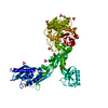 8f3fC 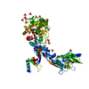 8f3gC 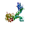 8f3hC 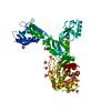 8f3iC 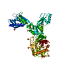 8f3jC 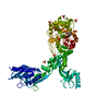 8f3lC 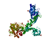 8f3mC 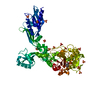 8f3nC 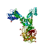 8f3oC 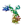 8f3pC 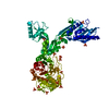 8f3qC 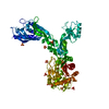 8f3rC 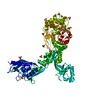 8f3sC 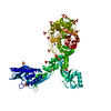 8f3tC 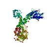 8f3uC 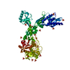 8f3zC  6mkaS C: citing same article ( S: Starting model for refinement |
|---|---|
| Similar structure data |
-Links
 PDBj
PDBj
- Assembly
| Deposited unit | 
| ||||||||
|---|---|---|---|---|---|---|---|---|---|
| 1 | 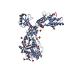
| ||||||||
| 2 | 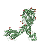
| ||||||||
| 3 | 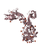
| ||||||||
| Unit cell |
|
-Components
| #1: Protein | Mass: 69858.781 Da / Num. of mol.: 3 Source method: isolated from a genetically manipulated source Source: (gene. exp.) Enterococcus faecium (bacteria) / Gene: pbp5 / Production host: #2: Chemical | ChemComp-SO4 /   Mass: 96.063 Da / Num. of mol.: 66 / Source method: obtained synthetically / Formula: SO4 Mass: 96.063 Da / Num. of mol.: 66 / Source method: obtained synthetically / Formula: SO4#3: Water | ChemComp-HOH / |  Mass: 18.015 Da / Num. of mol.: 128 / Source method: isolated from a natural source / Formula: H2O Mass: 18.015 Da / Num. of mol.: 128 / Source method: isolated from a natural source / Formula: H2OHas ligand of interest | N | |
|---|
-Experimental details
-Experiment
| Experiment | Method: X-RAY DIFFRACTION / Number of used crystals: 1 |
|---|
- Sample preparation
| Crystal | Density Matthews: 5.9 Å3/Da / Density % sol: 79.15 % |
|---|---|
| Crystal grow | Temperature: 298 K / Method: vapor diffusion, hanging drop / pH: 6 / Details: 0.1 M MES pH 6.0, 3.0 M ammonium sulfate |
-Data collection
| Diffraction | Mean temperature: 100 K / Serial crystal experiment: N |
|---|---|
| Diffraction source | Source: SYNCHROTRON / Site: SSRL / Beamline: BL12-2 / Wavelength: 0.97946 Å |
| Detector | Type: DECTRIS PILATUS 6M / Detector: PIXEL / Date: Mar 20, 2019 |
| Radiation | Protocol: SINGLE WAVELENGTH / Monochromatic (M) / Laue (L): M / Scattering type: x-ray |
| Radiation wavelength | Wavelength: 0.97946 Å / Relative weight: 1 |
| Reflection | Resolution: 3.59→39.63 Å / Num. obs: 57949 / % possible obs: 99.3 % / Redundancy: 6.8 % / CC1/2: 0.977 / Rmerge(I) obs: 0.293 / Rpim(I) all: 0.121 / Rrim(I) all: 0.317 / Χ2: 0.97 / Net I/σ(I): 8 / Num. measured all: 393565 |
| Reflection shell | Resolution: 3.59→3.68 Å / % possible obs: 92.8 % / Redundancy: 6.3 % / Rmerge(I) obs: 1.096 / Num. measured all: 26257 / Num. unique obs: 4185 / CC1/2: 0.605 / Rpim(I) all: 0.47 / Rrim(I) all: 1.197 / Χ2: 1.01 / Net I/σ(I) obs: 3.5 |
- Processing
| Software |
| ||||||||||||||||||||||||||||||||||||||||||||||||||||||||||||||||||||||||||||||||||||||||||||||||||||||||||||||||||||||||||||||||||||||||||||||||||||||||||
|---|---|---|---|---|---|---|---|---|---|---|---|---|---|---|---|---|---|---|---|---|---|---|---|---|---|---|---|---|---|---|---|---|---|---|---|---|---|---|---|---|---|---|---|---|---|---|---|---|---|---|---|---|---|---|---|---|---|---|---|---|---|---|---|---|---|---|---|---|---|---|---|---|---|---|---|---|---|---|---|---|---|---|---|---|---|---|---|---|---|---|---|---|---|---|---|---|---|---|---|---|---|---|---|---|---|---|---|---|---|---|---|---|---|---|---|---|---|---|---|---|---|---|---|---|---|---|---|---|---|---|---|---|---|---|---|---|---|---|---|---|---|---|---|---|---|---|---|---|---|---|---|---|---|---|---|
| Refinement | Method to determine structure: MOLECULAR REPLACEMENT Starting model: 6MKA Resolution: 3.59→39.63 Å / SU ML: 0.31 / Cross valid method: THROUGHOUT / σ(F): 1.34 / Phase error: 19.91 / Stereochemistry target values: ML
| ||||||||||||||||||||||||||||||||||||||||||||||||||||||||||||||||||||||||||||||||||||||||||||||||||||||||||||||||||||||||||||||||||||||||||||||||||||||||||
| Solvent computation | Shrinkage radii: 0.9 Å / VDW probe radii: 1.11 Å / Solvent model: FLAT BULK SOLVENT MODEL | ||||||||||||||||||||||||||||||||||||||||||||||||||||||||||||||||||||||||||||||||||||||||||||||||||||||||||||||||||||||||||||||||||||||||||||||||||||||||||
| Refinement step | Cycle: LAST / Resolution: 3.59→39.63 Å
| ||||||||||||||||||||||||||||||||||||||||||||||||||||||||||||||||||||||||||||||||||||||||||||||||||||||||||||||||||||||||||||||||||||||||||||||||||||||||||
| Refine LS restraints |
| ||||||||||||||||||||||||||||||||||||||||||||||||||||||||||||||||||||||||||||||||||||||||||||||||||||||||||||||||||||||||||||||||||||||||||||||||||||||||||
| LS refinement shell |
|
