+ データを開く
- 基本情報
基本情報
| 登録情報 | データベース: PDB / ID: 8f10 | ||||||||||||
|---|---|---|---|---|---|---|---|---|---|---|---|---|---|
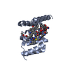
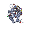
| タイトル | Structure of the MDM2 P53 binding domain in complex with H102, an all-D Helicon Polypeptide | ||||||||||||
 要素 要素 |
| ||||||||||||
 キーワード キーワード | LIGASE / E3 ligase / D-peptide / stapled peptide | ||||||||||||
| 機能・相同性 |  機能・相同性情報 機能・相同性情報cellular response to vitamin B1 / response to formaldehyde / response to water-immersion restraint stress / response to ether / traversing start control point of mitotic cell cycle / fibroblast activation / atrial septum development / regulation of protein catabolic process at postsynapse, modulating synaptic transmission / Trafficking of AMPA receptors / receptor serine/threonine kinase binding ...cellular response to vitamin B1 / response to formaldehyde / response to water-immersion restraint stress / response to ether / traversing start control point of mitotic cell cycle / fibroblast activation / atrial septum development / regulation of protein catabolic process at postsynapse, modulating synaptic transmission / Trafficking of AMPA receptors / receptor serine/threonine kinase binding / peroxisome proliferator activated receptor binding / negative regulation of intrinsic apoptotic signaling pathway by p53 class mediator / positive regulation of vascular associated smooth muscle cell migration / negative regulation of protein processing / SUMO transferase activity / response to steroid hormone / NEDD8 ligase activity / AKT phosphorylates targets in the cytosol / response to iron ion / atrioventricular valve morphogenesis / endocardial cushion morphogenesis / cellular response to peptide hormone stimulus / ventricular septum development / positive regulation of muscle cell differentiation / regulation of postsynaptic neurotransmitter receptor internalization / SUMOylation of ubiquitinylation proteins / cardiac septum morphogenesis / blood vessel development / ligase activity / cellular response to alkaloid / Constitutive Signaling by AKT1 E17K in Cancer / regulation of protein catabolic process / negative regulation of signal transduction by p53 class mediator / negative regulation of DNA damage response, signal transduction by p53 class mediator / SUMOylation of transcription factors / response to magnesium ion / cellular response to UV-C / protein sumoylation / cellular response to actinomycin D / blood vessel remodeling / cellular response to estrogen stimulus / protein localization to nucleus / ribonucleoprotein complex binding / protein autoubiquitination / positive regulation of vascular associated smooth muscle cell proliferation / NPAS4 regulates expression of target genes / transcription repressor complex / positive regulation of mitotic cell cycle / regulation of heart rate / proteolysis involved in protein catabolic process / positive regulation of protein export from nucleus / ubiquitin binding / response to cocaine / DNA damage response, signal transduction by p53 class mediator / Stabilization of p53 / establishment of protein localization / Regulation of RUNX3 expression and activity / cellular response to gamma radiation / protein destabilization / Oncogene Induced Senescence / RING-type E3 ubiquitin transferase / Regulation of TP53 Activity through Methylation / cellular response to growth factor stimulus / response to toxic substance / centriolar satellite / cellular response to hydrogen peroxide / protein polyubiquitination / ubiquitin-protein transferase activity / disordered domain specific binding / endocytic vesicle membrane / p53 binding / ubiquitin protein ligase activity / Signaling by ALK fusions and activated point mutants / Regulation of TP53 Degradation / positive regulation of proteasomal ubiquitin-dependent protein catabolic process / negative regulation of neuron projection development / 5S rRNA binding / protein-containing complex assembly / ubiquitin-dependent protein catabolic process / Oxidative Stress Induced Senescence / cellular response to hypoxia / Regulation of TP53 Activity through Phosphorylation / amyloid fibril formation / proteasome-mediated ubiquitin-dependent protein catabolic process / regulation of cell cycle / Ub-specific processing proteases / postsynaptic density / protein ubiquitination / response to xenobiotic stimulus / protein domain specific binding / response to antibiotic / negative regulation of DNA-templated transcription / positive regulation of cell population proliferation / apoptotic process / ubiquitin protein ligase binding / positive regulation of gene expression / negative regulation of apoptotic process / nucleolus / glutamatergic synapse / enzyme binding 類似検索 - 分子機能 | ||||||||||||
| 生物種 |  Homo sapiens (ヒト) Homo sapiens (ヒト)synthetic construct (人工物) | ||||||||||||
| 手法 |  X線回折 / X線回折 /  シンクロトロン / シンクロトロン /  分子置換 / 解像度: 1.28 Å 分子置換 / 解像度: 1.28 Å | ||||||||||||
 データ登録者 データ登録者 | Li, K. / Callahan, A.J. / Travaline, T.L. / Tokareva, O.S. / Swiecicki, J.-M. / Verdine, G.L. / Pentelute, B.L. / McGee, J.H. | ||||||||||||
| 資金援助 |  米国, 米国,  ドイツ, 3件 ドイツ, 3件
| ||||||||||||
 引用 引用 |  ジャーナル: Chemrxiv / 年: 2023 ジャーナル: Chemrxiv / 年: 2023タイトル: Single-Shot Flow Synthesis of D-Proteins for Mirror-Image Phage Display 著者: Callahan, A.J. / Gandhesiri, S. / Travaline, T.L. / Lozano Salazar, L. / Hanna, S. / Lee, Y.-C. / Li, K. / Tokareva, O.S. / Swiecicki, J.-M. / Loas, A. / Verdine, G.L. / McGee, J.H. / Pentelute, B.L. | ||||||||||||
| 履歴 |
|
- 構造の表示
構造の表示
| 構造ビューア | 分子:  Molmil Molmil Jmol/JSmol Jmol/JSmol |
|---|
- ダウンロードとリンク
- ダウンロード
| PDBx/mmCIF形式 |  8f10.cif.gz 8f10.cif.gz | 90.8 KB | 表示 |  PDBx/mmCIF形式 PDBx/mmCIF形式 |
|---|---|---|---|---|
| PDB形式 |  pdb8f10.ent.gz pdb8f10.ent.gz | 67.7 KB | 表示 |  PDB形式 PDB形式 |
| PDBx/mmJSON形式 |  8f10.json.gz 8f10.json.gz | ツリー表示 |  PDBx/mmJSON形式 PDBx/mmJSON形式 | |
| その他 |  その他のダウンロード その他のダウンロード |
-検証レポート
| 文書・要旨 |  8f10_validation.pdf.gz 8f10_validation.pdf.gz | 479.4 KB | 表示 |  wwPDB検証レポート wwPDB検証レポート |
|---|---|---|---|---|
| 文書・詳細版 |  8f10_full_validation.pdf.gz 8f10_full_validation.pdf.gz | 480.4 KB | 表示 | |
| XML形式データ |  8f10_validation.xml.gz 8f10_validation.xml.gz | 8.2 KB | 表示 | |
| CIF形式データ |  8f10_validation.cif.gz 8f10_validation.cif.gz | 10.8 KB | 表示 | |
| アーカイブディレクトリ |  https://data.pdbj.org/pub/pdb/validation_reports/f1/8f10 https://data.pdbj.org/pub/pdb/validation_reports/f1/8f10 ftp://data.pdbj.org/pub/pdb/validation_reports/f1/8f10 ftp://data.pdbj.org/pub/pdb/validation_reports/f1/8f10 | HTTPS FTP |
-関連構造データ
| 関連構造データ |  8f0zC  8f12C  8f13C  8f14C  8f15C  8f16C  8f17C  3g03S S: 精密化の開始モデル C: 同じ文献を引用 ( |
|---|---|
| 類似構造データ | 類似検索 - 機能・相同性  F&H 検索 F&H 検索 |
- リンク
リンク
- 集合体
集合体
| 登録構造単位 | 
| ||||||||
|---|---|---|---|---|---|---|---|---|---|
| 1 |
| ||||||||
| 単位格子 |
|
- 要素
要素
-タンパク質 / Polypeptide(D) , 2種, 2分子 AB
| #1: タンパク質 | 分子量: 11099.000 Da / 分子数: 1 / 由来タイプ: 組換発現 / 由来: (組換発現)  Homo sapiens (ヒト) / 遺伝子: MDM2 / 発現宿主: Homo sapiens (ヒト) / 遺伝子: MDM2 / 発現宿主:  |
|---|---|
| #2: Polypeptide(D) | 分子量: 1966.156 Da / 分子数: 1 / 由来タイプ: 合成 / 由来: (合成) synthetic construct (人工物) |
-非ポリマー , 6種, 114分子 


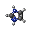
| #3: 化合物 | ChemComp-EDO / #4: 化合物 | ChemComp-CL / | #5: 化合物 | #6: 化合物 | ChemComp-WHL / | #7: 化合物 | ChemComp-IMD / | #8: 水 | ChemComp-HOH / | |
|---|
-詳細
| 研究の焦点であるリガンドがあるか | N |
|---|---|
| Has protein modification | Y |
-実験情報
-実験
| 実験 | 手法:  X線回折 / 使用した結晶の数: 1 X線回折 / 使用した結晶の数: 1 |
|---|
- 試料調製
試料調製
| 結晶 | マシュー密度: 1.81 Å3/Da / 溶媒含有率: 32.18 % |
|---|---|
| 結晶化 | 温度: 291 K / 手法: 蒸気拡散法, シッティングドロップ法 / 詳細: 0.01 M tri-Sodium citrate, 33 % w/v PEG 6000 |
-データ収集
| 回折 | 平均測定温度: 100 K / Serial crystal experiment: N |
|---|---|
| 放射光源 | 由来:  シンクロトロン / サイト: シンクロトロン / サイト:  NSLS-II NSLS-II  / ビームライン: 17-ID-2 / 波長: 0.97933 Å / ビームライン: 17-ID-2 / 波長: 0.97933 Å |
| 検出器 | タイプ: DECTRIS EIGER X 16M / 検出器: PIXEL / 日付: 2021年3月18日 |
| 放射 | プロトコル: SINGLE WAVELENGTH / 単色(M)・ラウエ(L): M / 散乱光タイプ: x-ray |
| 放射波長 | 波長: 0.97933 Å / 相対比: 1 |
| 反射 | 解像度: 1.28→42.17 Å / Num. obs: 20614 / % possible obs: 85.3 % / 冗長度: 4.8 % / Rmerge(I) obs: 0.049 / Net I/σ(I): 18.2 |
| 反射 シェル | 解像度: 1.28→1.3 Å / Rmerge(I) obs: 0.42 / Mean I/σ(I) obs: 2.1 / Num. unique obs: 233 |
- 解析
解析
| ソフトウェア |
| ||||||||||||||||||||||||||||||||||||||||||||||||||||||||
|---|---|---|---|---|---|---|---|---|---|---|---|---|---|---|---|---|---|---|---|---|---|---|---|---|---|---|---|---|---|---|---|---|---|---|---|---|---|---|---|---|---|---|---|---|---|---|---|---|---|---|---|---|---|---|---|---|---|
| 精密化 | 構造決定の手法:  分子置換 分子置換開始モデル: 3G03 解像度: 1.28→28.02 Å / SU ML: 0.12 / 交差検証法: THROUGHOUT / σ(F): 1.36 / 位相誤差: 21.14 / 立体化学のターゲット値: ML
| ||||||||||||||||||||||||||||||||||||||||||||||||||||||||
| 溶媒の処理 | 減衰半径: 0.9 Å / VDWプローブ半径: 1.11 Å / 溶媒モデル: FLAT BULK SOLVENT MODEL | ||||||||||||||||||||||||||||||||||||||||||||||||||||||||
| 原子変位パラメータ | Biso max: 75.25 Å2 / Biso mean: 18.4648 Å2 / Biso min: 7.86 Å2 | ||||||||||||||||||||||||||||||||||||||||||||||||||||||||
| 精密化ステップ | サイクル: final / 解像度: 1.28→28.02 Å
| ||||||||||||||||||||||||||||||||||||||||||||||||||||||||
| LS精密化 シェル | Refine-ID: X-RAY DIFFRACTION / Rfactor Rfree error: 0 / Total num. of bins used: 7
| ||||||||||||||||||||||||||||||||||||||||||||||||||||||||
| 精密化 TLS | 手法: refined / Origin x: -0.7968 Å / Origin y: 5.4555 Å / Origin z: -11.0623 Å
| ||||||||||||||||||||||||||||||||||||||||||||||||||||||||
| 精密化 TLSグループ |
|
 ムービー
ムービー コントローラー


 PDBj
PDBj


