 Movie
Movie Controller

[English] 日本語
 Yorodumi
- PDB-8f0z: Structure of the MDM2 P53 binding domain in complex with H101, an... -
+ Open data

- Basic information
| Entry | Database: PDB / ID: 8f0z | ||||||||||||
|---|---|---|---|---|---|---|---|---|---|---|---|---|---|
| Title | Structure of the MDM2 P53 binding domain in complex with H101, an all-D Helicon Polypeptide | ||||||||||||
 Components Components |
| ||||||||||||
 Keywords Keywords | LIGASE / E3 ligase / D-peptide / stapled peptide | ||||||||||||
| Function / homology |  Function and homology information Function and homology informationcellular response to vitamin B1 / response to formaldehyde / response to water-immersion restraint stress / response to ether / traversing start control point of mitotic cell cycle / atrial septum development / fibroblast activation / regulation of protein catabolic process at postsynapse, modulating synaptic transmission / Trafficking of AMPA receptors / negative regulation of intrinsic apoptotic signaling pathway by p53 class mediator ...cellular response to vitamin B1 / response to formaldehyde / response to water-immersion restraint stress / response to ether / traversing start control point of mitotic cell cycle / atrial septum development / fibroblast activation / regulation of protein catabolic process at postsynapse, modulating synaptic transmission / Trafficking of AMPA receptors / negative regulation of intrinsic apoptotic signaling pathway by p53 class mediator / receptor serine/threonine kinase binding / negative regulation of protein processing / response to steroid hormone / SUMO transferase activity / peroxisome proliferator activated receptor binding / positive regulation of vascular associated smooth muscle cell migration / atrioventricular valve morphogenesis / AKT phosphorylates targets in the cytosol / response to iron ion / NEDD8 ligase activity / endocardial cushion morphogenesis / ventricular septum development / cellular response to peptide hormone stimulus / positive regulation of muscle cell differentiation / cardiac septum morphogenesis / regulation of postsynaptic neurotransmitter receptor internalization / SUMOylation of ubiquitinylation proteins / cellular response to alkaloid / blood vessel development / ligase activity / Constitutive Signaling by AKT1 E17K in Cancer / negative regulation of DNA damage response, signal transduction by p53 class mediator / cellular response to antibiotic / SUMOylation of transcription factors / negative regulation of signal transduction by p53 class mediator / regulation of protein catabolic process / cellular response to UV-C / cellular response to estrogen stimulus / response to magnesium ion / protein sumoylation / blood vessel remodeling / ribonucleoprotein complex binding / protein localization to nucleus / protein autoubiquitination / positive regulation of vascular associated smooth muscle cell proliferation / NPAS4 regulates expression of target genes / transcription repressor complex / positive regulation of mitotic cell cycle / regulation of heart rate / : / positive regulation of protein export from nucleus / response to cocaine / ubiquitin binding / DNA damage response, signal transduction by p53 class mediator / establishment of protein localization / sperm end piece / Stabilization of p53 / cellular response to gamma radiation / Regulation of RUNX3 expression and activity / RING-type E3 ubiquitin transferase / Oncogene Induced Senescence / Regulation of TP53 Activity through Methylation / protein destabilization / cellular response to growth factor stimulus / Degradation of CDH1 / response to toxic substance / cellular response to hydrogen peroxide / centriolar satellite / disordered domain specific binding / protein polyubiquitination / p53 binding / ubiquitin-protein transferase activity / endocytic vesicle membrane / Signaling by ALK fusions and activated point mutants / Regulation of TP53 Degradation / ubiquitin protein ligase activity / positive regulation of proteasomal ubiquitin-dependent protein catabolic process / negative regulation of neuron projection development / sperm principal piece / 5S rRNA binding / protein-containing complex assembly / sperm midpiece / Oxidative Stress Induced Senescence / cellular response to hypoxia / Regulation of TP53 Activity through Phosphorylation / amyloid fibril formation / ubiquitin-dependent protein catabolic process / proteasome-mediated ubiquitin-dependent protein catabolic process / regulation of cell cycle / postsynaptic density / Ub-specific processing proteases / protein ubiquitination / response to xenobiotic stimulus / protein domain specific binding / response to antibiotic / negative regulation of DNA-templated transcription / apoptotic process / positive regulation of cell population proliferation / ubiquitin protein ligase binding / positive regulation of gene expression Similarity search - Function | ||||||||||||
| Biological species |  Homo sapiens (human) Homo sapiens (human)synthetic construct (others) | ||||||||||||
| Method |  X-RAY DIFFRACTION / SYNCHROTRON / MOLECULAR REPLACEMENT / Resolution: 1.61 Å X-RAY DIFFRACTION / SYNCHROTRON / MOLECULAR REPLACEMENT / Resolution: 1.61 Å | ||||||||||||
 Authors Authors | Li, K. / Callahan, A.J. / Travaline, T.L. / Tokareva, O.S. / Swiecicki, J.-M. / Verdine, G.L. / Pentelute, B.L. / McGee, J.H. | ||||||||||||
| Funding support |  United States, United States,  Germany, 3items Germany, 3items
| ||||||||||||
 Citation Citation | Journal: Chemrxiv / Year: 2023 Title: Single-Shot Flow Synthesis of D-Proteins for Mirror-Image Phage Display Authors: Callahan, A.J. / Gandhesiri, S. / Travaline, T.L. / Lozano Salazar, L. / Hanna, S. / Lee, Y.-C. / Li, K. / Tokareva, O.S. / Swiecicki, J.-M. / Loas, A. / Verdine, G.L. / McGee, J.H. / Pentelute, B.L. | ||||||||||||
| History |
|
- Structure visualization
| Structure viewer | Molecule: MolmilJmol/JSmol |
|---|
- Downloads & links
-Download
| PDBx/mmCIF format | 8f0z.cif.gz | 42.5 KB | Display | PDBx/mmCIF format |
|---|---|---|---|---|
| PDB format | pdb8f0z.ent.gz | 26.1 KB | Display | PDB format |
| PDBx/mmJSON format | 8f0z.json.gz | Tree view | PDBx/mmJSON format | |
| Others |  Other downloads Other downloads |
-Validation report
| Arichive directory | https://data.pdbj.org/pub/pdb/validation_reports/f0/8f0zftp://data.pdbj.org/pub/pdb/validation_reports/f0/8f0z | HTTPS FTP |
|---|
-Related structure data
| Related structure data |  8f10C  8f12C  8f13C  8f14C  8f15C  8f16C 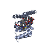 8f17C  3g03S S: Starting model for refinement C: citing same article ( |
|---|---|
| Similar structure data |
-Links
 PDBj
PDBj


- Assembly
| Deposited unit | 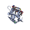
| |||||||||
|---|---|---|---|---|---|---|---|---|---|---|
| 1 |
| |||||||||
| Unit cell |
| |||||||||
| Components on special symmetry positions |
|
-Components
| #1: Protein | Mass: 11099.000 Da / Num. of mol.: 1 Source method: isolated from a genetically manipulated source Source: (gene. exp.) Homo sapiens (human) / Gene: MDM2 / Production host:  References: UniProt: Q00987, RING-type E3 ubiquitin transferase |
|---|---|
| #2: Polypeptide(D) | Mass: 1999.189 Da / Num. of mol.: 1 / Source method: obtained synthetically / Source: (synth.) synthetic construct (others) |
| #3: Chemical | ChemComp-CL /   Mass: 35.453 Da / Num. of mol.: 1 / Source method: obtained synthetically / Formula: Cl Mass: 35.453 Da / Num. of mol.: 1 / Source method: obtained synthetically / Formula: Cl |
| #4: Chemical | ChemComp-WHL /   Mass: 192.214 Da / Num. of mol.: 1 / Source method: obtained synthetically / Formula: C10H12N2O2 Mass: 192.214 Da / Num. of mol.: 1 / Source method: obtained synthetically / Formula: C10H12N2O2 |
| #5: Water | ChemComp-HOH /  Mass: 18.015 Da / Num. of mol.: 67 / Source method: isolated from a natural source / Formula: H2O Mass: 18.015 Da / Num. of mol.: 67 / Source method: isolated from a natural source / Formula: H2O |
| Has ligand of interest | N |
| Has protein modification | Y |
-Experimental details
-Experiment
| Experiment | Method: X-RAY DIFFRACTION / Number of used crystals: 1 |
|---|
- Sample preparation
| Crystal | Density Matthews: 2.22 Å3/Da / Density % sol: 44.59 % |
|---|---|
| Crystal grow | Temperature: 291 K / Method: vapor diffusion, sitting drop / pH: 7 / Details: 3.5 M Sodium Formate pH 7.0 |
-Data collection
| Diffraction | Mean temperature: 100 K / Serial crystal experiment: N |
|---|---|
| Diffraction source | Source: SYNCHROTRON / Site: APS / Beamline: 23-ID-B / Wavelength: 1.03319 Å |
| Detector | Type: DECTRIS EIGER X 16M / Detector: PIXEL / Date: Mar 31, 2021 |
| Radiation | Protocol: SINGLE WAVELENGTH / Monochromatic (M) / Laue (L): M / Scattering type: x-ray |
| Radiation wavelength | Wavelength: 1.03319 Å / Relative weight: 1 |
| Reflection | Resolution: 1.61→40.87 Å / Num. obs: 15696 / % possible obs: 99.6 % / Redundancy: 12.8 % / CC1/2: 0.998 / Rmerge(I) obs: 0.067 / Net I/σ(I): 19.3 |
| Reflection shell | Resolution: 1.61→1.64 Å / Redundancy: 12.9 % / Mean I/σ(I) obs: 2.1 / Num. unique obs: 774 / CC1/2: 0.774 / % possible all: 100 |
- Processing
| Software |
| ||||||||||||||||||
|---|---|---|---|---|---|---|---|---|---|---|---|---|---|---|---|---|---|---|---|
| Refinement | Method to determine structure: MOLECULAR REPLACEMENT Starting model: 3G03 Resolution: 1.61→40.87 Å / Cross valid method: THROUGHOUT
| ||||||||||||||||||
| Displacement parameters | Biso max: 95.8 Å2 / Biso mean: 35.5855 Å2 / Biso min: 18.36 Å2 | ||||||||||||||||||
| Refinement step | Cycle: LAST / Resolution: 1.61→40.87 Å
|
