 Movie
Movie Controller


+ Open data

- Basic information
| Entry | Database: PDB / ID: 8f0m | |||||||||||||||||||||
|---|---|---|---|---|---|---|---|---|---|---|---|---|---|---|---|---|---|---|---|---|---|---|
| Title | Monobody 12D5 bound to KRAS(G12D) | |||||||||||||||||||||
 Components Components |
| |||||||||||||||||||||
 Keywords Keywords | SIGNALING PROTEIN/DE NOVO PROTEIN / SIGNALING PROTEIN-DE NOVO PROTEIN complex | |||||||||||||||||||||
| Function / homology |  Function and homology information Function and homology information | |||||||||||||||||||||
| Biological species |  Homo sapiens (human) Homo sapiens (human) | |||||||||||||||||||||
| Method |  X-RAY DIFFRACTION / SYNCHROTRON / MOLECULAR REPLACEMENT / Resolution: 2.44 Å X-RAY DIFFRACTION / SYNCHROTRON / MOLECULAR REPLACEMENT / Resolution: 2.44 Å | |||||||||||||||||||||
 Authors Authors | Hattori, T. / Glasser, E. / Akkapeddi, P. / Ketavarapu, G. / Teng, K.W. / Koide, A. / Koide, S. | |||||||||||||||||||||
| Funding support |  United States, 6items United States, 6items
| |||||||||||||||||||||
 Citation Citation | Journal: Proc.Natl.Acad.Sci.USA / Year: 2023 Title: Exploring switch II pocket conformation of KRAS(G12D) with mutant-selective monobody inhibitors. Authors: Akkapeddi, P. / Hattori, T. / Khan, I. / Glasser, E. / Koide, A. / Ketavarapu, G. / Whaby, M. / Zuberi, M. / Teng, K.W. / Lefler, J. / Maso, L. / Bang, I. / Ostrowski, M.C. / O'Bryan, J.P. / Koide, S. | |||||||||||||||||||||
| History |
|
- Structure visualization
| Structure viewer | Molecule: MolmilJmol/JSmol |
|---|
- Downloads & links
-Download
| PDBx/mmCIF format | 8f0m.cif.gz | 225.3 KB | Display | PDBx/mmCIF format |
|---|---|---|---|---|
| PDB format | pdb8f0m.ent.gz | 176.9 KB | Display | PDB format |
| PDBx/mmJSON format | 8f0m.json.gz | Tree view | PDBx/mmJSON format | |
| Others |  Other downloads Other downloads |
-Validation report
| Summary document | 8f0m_validation.pdf.gz | 1.1 MB | Display | wwPDB validaton report |
|---|---|---|---|---|
| Full document | 8f0m_full_validation.pdf.gz | 1.1 MB | Display | |
| Data in XML | 8f0m_validation.xml.gz | 21.6 KB | Display | |
| Data in CIF | 8f0m_validation.cif.gz | 29.3 KB | Display | |
| Arichive directory | https://data.pdbj.org/pub/pdb/validation_reports/f0/8f0mftp://data.pdbj.org/pub/pdb/validation_reports/f0/8f0m | HTTPS FTP |
-Related structure data
| Related structure data | 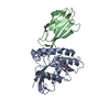 8ezgC 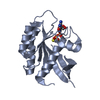 5vpzS  7l0gS C: citing same article ( S: Starting model for refinement |
|---|---|
| Similar structure data |
-Links
 PDBj
PDBj


- Assembly
| Deposited unit | 
| ||||||||
|---|---|---|---|---|---|---|---|---|---|
| 1 | 
| ||||||||
| 2 | 
| ||||||||
| Unit cell |
|
-Components
-Protein / Antibody / Sugars , 3 types, 6 molecules ACBD
| #1: Protein | Mass: 19451.809 Da / Num. of mol.: 2 / Mutation: G12D, C51S, C80L, C118S Source method: isolated from a genetically manipulated source Source: (gene. exp.) Homo sapiens (human) / Gene: KRAS, KRAS2, RASK2 / Production host:  #2: Antibody | Mass: 10311.420 Da / Num. of mol.: 2 Source method: isolated from a genetically manipulated source Source: (gene. exp.) Homo sapiens (human) / Production host: #5: Sugar |  Type: D-saccharide, alpha linking / Mass: 180.156 Da / Num. of mol.: 2 / Source method: obtained synthetically / Formula: C6H12O6 Type: D-saccharide, alpha linking / Mass: 180.156 Da / Num. of mol.: 2 / Source method: obtained synthetically / Formula: C6H12O6 |
|---|
-Non-polymers , 4 types, 106 molecules 


| #3: Chemical |  Mass: 24.305 Da / Num. of mol.: 2 / Source method: obtained synthetically / Formula: Mg Mass: 24.305 Da / Num. of mol.: 2 / Source method: obtained synthetically / Formula: Mg#4: Chemical | ChemComp-GDP / |  Type: RNA linking / Mass: 443.201 Da / Num. of mol.: 1 / Source method: obtained synthetically / Formula: C10H15N5O11P2 / Feature type: SUBJECT OF INVESTIGATION / Comment: GDP, energy-carrying molecule*YM Type: RNA linking / Mass: 443.201 Da / Num. of mol.: 1 / Source method: obtained synthetically / Formula: C10H15N5O11P2 / Feature type: SUBJECT OF INVESTIGATION / Comment: GDP, energy-carrying molecule*YM#6: Chemical | ChemComp-GSP / |  Mass: 539.246 Da / Num. of mol.: 1 / Source method: isolated from a natural source / Formula: C10H16N5O13P3S / Feature type: SUBJECT OF INVESTIGATION Mass: 539.246 Da / Num. of mol.: 1 / Source method: isolated from a natural source / Formula: C10H16N5O13P3S / Feature type: SUBJECT OF INVESTIGATION#7: Water | ChemComp-HOH / | Mass: 18.015 Da / Num. of mol.: 102 / Source method: isolated from a natural source / Formula: H2O |
|---|
-Details
| Has ligand of interest | Y |
|---|
-Experimental details
-Experiment
| Experiment | Method: X-RAY DIFFRACTION / Number of used crystals: 1 |
|---|
- Sample preparation
| Crystal | Density Matthews: 2.56 Å3/Da / Density % sol: 52.02 % |
|---|---|
| Crystal grow | Temperature: 291 K / Method: vapor diffusion, sitting drop Details: 0.2 M sodium sulfate decahydrate, 20% w/v PEG3350, 0.05% w/v benzamidine hydrochloride |
-Data collection
| Diffraction | Mean temperature: 100 K / Serial crystal experiment: N |
|---|---|
| Diffraction source | Source: SYNCHROTRON / Site: APS / Beamline: 19-ID / Wavelength: 0.97918 Å |
| Detector | Type: DECTRIS PILATUS3 6M / Detector: PIXEL / Date: Sep 24, 2022 |
| Radiation | Monochromator: Si(111) / Protocol: SINGLE WAVELENGTH / Monochromatic (M) / Laue (L): M / Scattering type: x-ray |
| Radiation wavelength | Wavelength: 0.97918 Å / Relative weight: 1 |
| Reflection | Resolution: 2.44→50 Å / Num. obs: 23370 / % possible obs: 98.4 % / Redundancy: 3.3 % / CC1/2: 0.939 / CC star: 0.984 / Rmerge(I) obs: 0.091 / Rpim(I) all: 0.061 / Rrim(I) all: 0.11 / Χ2: 1.043 / Net I/σ(I): 12.4 |
| Reflection shell | Resolution: 2.44→2.48 Å / Redundancy: 3.2 % / Rmerge(I) obs: 0.308 / Mean I/σ(I) obs: 5 / Num. unique obs: 1168 / CC1/2: 0.905 / CC star: 0.975 / Rpim(I) all: 0.207 / Rrim(I) all: 0.373 / Χ2: 1.024 / % possible all: 99.7 |
- Processing
| Software |
| |||||||||||||||||||||||||||||||||||||||||||||||||||||||||||||||
|---|---|---|---|---|---|---|---|---|---|---|---|---|---|---|---|---|---|---|---|---|---|---|---|---|---|---|---|---|---|---|---|---|---|---|---|---|---|---|---|---|---|---|---|---|---|---|---|---|---|---|---|---|---|---|---|---|---|---|---|---|---|---|---|---|
| Refinement | Method to determine structure: MOLECULAR REPLACEMENT Starting model: PDB entries 7L0G & 5VPZ Resolution: 2.44→42.9 Å / SU ML: 0.23 / Cross valid method: THROUGHOUT / σ(F): 1.36 / Phase error: 25.14 / Stereochemistry target values: ML
| |||||||||||||||||||||||||||||||||||||||||||||||||||||||||||||||
| Solvent computation | Shrinkage radii: 0.9 Å / VDW probe radii: 1.11 Å / Solvent model: FLAT BULK SOLVENT MODEL | |||||||||||||||||||||||||||||||||||||||||||||||||||||||||||||||
| Refinement step | Cycle: LAST / Resolution: 2.44→42.9 Å
| |||||||||||||||||||||||||||||||||||||||||||||||||||||||||||||||
| Refine LS restraints |
| |||||||||||||||||||||||||||||||||||||||||||||||||||||||||||||||
| LS refinement shell |
| |||||||||||||||||||||||||||||||||||||||||||||||||||||||||||||||
| Refinement TLS params. | Method: refined / Origin x: 22.1947 Å / Origin y: 29.3203 Å / Origin z: 36.6075 Å
| |||||||||||||||||||||||||||||||||||||||||||||||||||||||||||||||
| Refinement TLS group | Selection details: all |
