 Movie
Movie Controller

[English] 日本語
 Yorodumi
- PDB-8eo6: Crystal structure of metagenomic class A beta-lactamase precursor... -
+ Open data

- Basic information
| Entry | Database: PDB / ID: 8eo6 | |||||||||
|---|---|---|---|---|---|---|---|---|---|---|
| Title | Crystal structure of metagenomic class A beta-lactamase precursor LRA-5 in complex with ceftazidime at 2.35 Angstrom resolution | |||||||||
 Components Components | LRA-5 | |||||||||
 Keywords Keywords | ANTIMICROBIAL PROTEIN / beta-lactamase precursor / environmental resistome / soil metagenome / ceftazidime / oxyimino-cephalosporin | |||||||||
| Function / homology | Beta-lactamase class A, catalytic domain / Beta-lactamase enzyme family / beta-lactam antibiotic catabolic process / Beta-lactamase, class-A / Beta-lactamase/transpeptidase-like / beta-lactamase activity / response to antibiotic / ACYLATED CEFTAZIDIME / LRA-5 Function and homology information Function and homology information | |||||||||
| Biological species |  uncultured soil bacterium (environmental samples) uncultured soil bacterium (environmental samples) | |||||||||
| Method |  X-RAY DIFFRACTION / SYNCHROTRON / MOLECULAR REPLACEMENT / Resolution: 2.35 Å X-RAY DIFFRACTION / SYNCHROTRON / MOLECULAR REPLACEMENT / Resolution: 2.35 Å | |||||||||
 Authors Authors | Power, P. / D'Amico Gonzalez, G. / Centron, D. / Gutkind, G. / Handelsman, J. / Klinke, S. | |||||||||
| Funding support |  Argentina, 2items Argentina, 2items
| |||||||||
 Citation Citation | Journal: to be published Title: Playing beta-Lactamase Evolution: Metagenomic Class A beta-Lactamase LRA-5 is an Inactive Enzyme Capable of Rendering an Active beta-Lactamase by Introduction of Y69Q and V166E Substitutions Authors: D'Amico Gonzalez, G. / Handelsman, J. / Centron, D. / Gutkind, G. / Klinke, S. / Power, P. | |||||||||
| History |
|
- Structure visualization
| Structure viewer | Molecule: MolmilJmol/JSmol |
|---|
- Downloads & links
-Download
| PDBx/mmCIF format | 8eo6.cif.gz | 115.3 KB | Display | PDBx/mmCIF format |
|---|---|---|---|---|
| PDB format | pdb8eo6.ent.gz | 87.6 KB | Display | PDB format |
| PDBx/mmJSON format | 8eo6.json.gz | Tree view | PDBx/mmJSON format | |
| Others |  Other downloads Other downloads |
-Validation report
| Arichive directory | https://data.pdbj.org/pub/pdb/validation_reports/eo/8eo6ftp://data.pdbj.org/pub/pdb/validation_reports/eo/8eo6 | HTTPS FTP |
|---|
-Related structure data
| Related structure data | 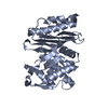 8eo7C 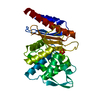 1e25S S: Starting model for refinement C: citing same article ( |
|---|---|
| Similar structure data |
-Links
 PDBj
PDBj


- Assembly
| Deposited unit | 
| ||||||||
|---|---|---|---|---|---|---|---|---|---|
| 1 | 
| ||||||||
| 2 | 
| ||||||||
| Unit cell |
|
-Components
| #1: Protein | Mass: 30977.408 Da / Num. of mol.: 2 Source method: isolated from a genetically manipulated source Source: (gene. exp.) uncultured soil bacterium (environmental samples)Gene: blaLRA-5, AKSOIL_0013 / Plasmid: pET28a / Production host: #2: Chemical | 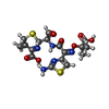  Mass: 469.492 Da / Num. of mol.: 2 / Source method: obtained synthetically / Formula: C17H19N5O7S2 / Feature type: SUBJECT OF INVESTIGATION Mass: 469.492 Da / Num. of mol.: 2 / Source method: obtained synthetically / Formula: C17H19N5O7S2 / Feature type: SUBJECT OF INVESTIGATION#3: Water | ChemComp-HOH / |  Mass: 18.015 Da / Num. of mol.: 42 / Source method: isolated from a natural source / Formula: H2O Mass: 18.015 Da / Num. of mol.: 42 / Source method: isolated from a natural source / Formula: H2OHas ligand of interest | Y | Has protein modification | Y | |
|---|
-Experimental details
-Experiment
| Experiment | Method: X-RAY DIFFRACTION / Number of used crystals: 1 |
|---|
- Sample preparation
| Crystal | Density Matthews: 3.41 Å3/Da / Density % sol: 64 % / Description: long bars |
|---|---|
| Crystal grow | Temperature: 294 K / Method: vapor diffusion, hanging drop / pH: 10.5 / Details: 1.6 M sodium formate |
-Data collection
| Diffraction | Mean temperature: 100 K / Serial crystal experiment: N |
|---|---|
| Diffraction source | Source: SYNCHROTRON / Site: SOLEIL  / Beamline: PROXIMA 2 / Wavelength: 0.980112 Å / Beamline: PROXIMA 2 / Wavelength: 0.980112 Å |
| Detector | Type: DECTRIS EIGER X 9M / Detector: PIXEL / Date: Oct 16, 2021 Details: convex prefocusing mirror and a Kirkpatrick-Baez pair of focusing mirrors |
| Radiation | Monochromator: Cryogenically cooled channel cut crystal monochromator Protocol: SINGLE WAVELENGTH / Monochromatic (M) / Laue (L): M / Scattering type: x-ray |
| Radiation wavelength | Wavelength: 0.980112 Å / Relative weight: 1 |
| Reflection | Resolution: 2.35→43.66 Å / Num. obs: 36200 / % possible obs: 99.7 % / Redundancy: 13.4 % / CC1/2: 0.997 / Rrim(I) all: 0.283 / Net I/σ(I): 11.78 |
| Reflection shell | Resolution: 2.35→2.49 Å / Redundancy: 12.72 % / Mean I/σ(I) obs: 1.62 / Num. unique obs: 5654 / CC1/2: 0.495 / Rrim(I) all: 2.33 / % possible all: 98.1 |
- Processing
| Software |
| ||||||||||||||||||||||||||||||||||||||||||||||||||||||||||||||||||||||||||||||||||||||||||||||||||
|---|---|---|---|---|---|---|---|---|---|---|---|---|---|---|---|---|---|---|---|---|---|---|---|---|---|---|---|---|---|---|---|---|---|---|---|---|---|---|---|---|---|---|---|---|---|---|---|---|---|---|---|---|---|---|---|---|---|---|---|---|---|---|---|---|---|---|---|---|---|---|---|---|---|---|---|---|---|---|---|---|---|---|---|---|---|---|---|---|---|---|---|---|---|---|---|---|---|---|---|
| Refinement | Method to determine structure: MOLECULAR REPLACEMENT / Starting model: 1.0E+25 / Resolution: 2.35→43.66 Å / SU ML: 0.3 / Cross valid method: THROUGHOUT / σ(F): 1.33 / Phase error: 22.04 / Stereochemistry target values: ML
| ||||||||||||||||||||||||||||||||||||||||||||||||||||||||||||||||||||||||||||||||||||||||||||||||||
| Solvent computation | Shrinkage radii: 0.9 Å / VDW probe radii: 1.1 Å / Solvent model: FLAT BULK SOLVENT MODEL | ||||||||||||||||||||||||||||||||||||||||||||||||||||||||||||||||||||||||||||||||||||||||||||||||||
| Displacement parameters | Biso mean: 46 Å2 | ||||||||||||||||||||||||||||||||||||||||||||||||||||||||||||||||||||||||||||||||||||||||||||||||||
| Refinement step | Cycle: LAST / Resolution: 2.35→43.66 Å
| ||||||||||||||||||||||||||||||||||||||||||||||||||||||||||||||||||||||||||||||||||||||||||||||||||
| Refine LS restraints |
| ||||||||||||||||||||||||||||||||||||||||||||||||||||||||||||||||||||||||||||||||||||||||||||||||||
| LS refinement shell |
|
