 Movie
Movie Controller

[English] 日本語
 Yorodumi
- PDB-8ehe: Structure of Tannerella forsythia potempin C in complex with mirolase -
+ Open data

- Basic information
| Entry | Database: PDB / ID: 8ehe | ||||||
|---|---|---|---|---|---|---|---|
| Title | Structure of Tannerella forsythia potempin C in complex with mirolase | ||||||
 Components Components |
| ||||||
 Keywords Keywords | HYDROLASE INHIBITOR / Serine-peptidase inhibitor / HYDROLASE | ||||||
| Function / homology |  Function and homology information Function and homology informationprotein processing / serine-type endopeptidase activity / membrane / metal ion binding Similarity search - Function | ||||||
| Biological species |  Tannerella forsythia (bacteria) Tannerella forsythia (bacteria) | ||||||
| Method |  X-RAY DIFFRACTION / SYNCHROTRON / MOLECULAR REPLACEMENT / Resolution: 1.1 Å X-RAY DIFFRACTION / SYNCHROTRON / MOLECULAR REPLACEMENT / Resolution: 1.1 Å | ||||||
 Authors Authors | Gomis-Ruth, F.X. | ||||||
| Funding support | 1items
| ||||||
 Citation Citation | Journal: Chem Sci / Year: 2023 Title: A unique network of attack, defence and competence on the outer membrane of the periodontitis pathogen Tannerella forsythia. Authors: Ksiazek, M. / Goulas, T. / Mizgalska, D. / Rodriguez-Banqueri, A. / Eckhard, U. / Veillard, F. / Waligorska, I. / Benedyk-Machaczka, M. / Sochaj-Gregorczyk, A.M. / Madej, M. / Thogersen, I.B. ...Authors: Ksiazek, M. / Goulas, T. / Mizgalska, D. / Rodriguez-Banqueri, A. / Eckhard, U. / Veillard, F. / Waligorska, I. / Benedyk-Machaczka, M. / Sochaj-Gregorczyk, A.M. / Madej, M. / Thogersen, I.B. / Enghild, J.J. / Cuppari, A. / Arolas, J.L. / de Diego, I. / Lopez-Pelegrin, M. / Garcia-Ferrer, I. / Guevara, T. / Dive, V. / Zani, M.L. / Moreau, T. / Potempa, J. / Gomis-Ruth, F.X. | ||||||
| History |
|
- Structure visualization
| Structure viewer | Molecule: MolmilJmol/JSmol |
|---|
- Downloads & links
-Download
| PDBx/mmCIF format | 8ehe.cif.gz | 379 KB | Display | PDBx/mmCIF format |
|---|---|---|---|---|
| PDB format | pdb8ehe.ent.gz | 307.9 KB | Display | PDB format |
| PDBx/mmJSON format | 8ehe.json.gz | Tree view | PDBx/mmJSON format | |
| Others |  Other downloads Other downloads |
-Validation report
| Arichive directory | https://data.pdbj.org/pub/pdb/validation_reports/eh/8eheftp://data.pdbj.org/pub/pdb/validation_reports/eh/8ehe | HTTPS FTP |
|---|
-Related structure data
| Related structure data | 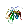 8b2mC  8b2nC 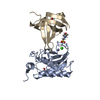 8b2qC 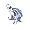 8ehbC 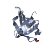 8ehcC 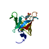 8ehdC 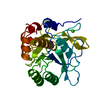 1st3S S: Starting model for refinement C: citing same article ( |
|---|---|
| Similar structure data |
-Links
 PDBj
PDBj


- Assembly
| Deposited unit | 
| ||||||||
|---|---|---|---|---|---|---|---|---|---|
| 1 |
| ||||||||
| Unit cell |
|
-Components
-Protein , 2 types, 2 molecules AB
| #1: Protein | Mass: 37594.961 Da / Num. of mol.: 1 Source method: isolated from a genetically manipulated source Source: (gene. exp.) Tannerella forsythia (bacteria) / Production host: |
|---|---|
| #2: Protein | Mass: 16974.893 Da / Num. of mol.: 1 Source method: isolated from a genetically manipulated source Source: (gene. exp.) Tannerella forsythia (bacteria) / Gene: CLI86_13365, TFUB20_02175 / Production host: |
-Non-polymers , 5 types, 769 molecules 


| #3: Chemical | ChemComp-NA /  Mass: 22.990 Da / Num. of mol.: 1 / Source method: obtained synthetically / Formula: Na / Feature type: SUBJECT OF INVESTIGATION Mass: 22.990 Da / Num. of mol.: 1 / Source method: obtained synthetically / Formula: Na / Feature type: SUBJECT OF INVESTIGATION | ||||||
|---|---|---|---|---|---|---|---|
| #4: Chemical | ChemComp-CA /  Mass: 40.078 Da / Num. of mol.: 6 / Source method: obtained synthetically / Formula: Ca / Feature type: SUBJECT OF INVESTIGATION Mass: 40.078 Da / Num. of mol.: 6 / Source method: obtained synthetically / Formula: Ca / Feature type: SUBJECT OF INVESTIGATION#5: Chemical | ChemComp-GOL /  Mass: 92.094 Da / Num. of mol.: 8 / Source method: obtained synthetically / Formula: C3H8O3 Mass: 92.094 Da / Num. of mol.: 8 / Source method: obtained synthetically / Formula: C3H8O3#6: Chemical |  Mass: 62.068 Da / Num. of mol.: 2 / Source method: obtained synthetically / Formula: C2H6O2 Mass: 62.068 Da / Num. of mol.: 2 / Source method: obtained synthetically / Formula: C2H6O2#7: Water | ChemComp-HOH / | Mass: 18.015 Da / Num. of mol.: 752 / Source method: isolated from a natural source / Formula: H2O |
-Details
| Has ligand of interest | Y |
|---|---|
| Has protein modification | Y |
-Experimental details
-Experiment
| Experiment | Method: X-RAY DIFFRACTION / Number of used crystals: 1 |
|---|
- Sample preparation
| Crystal | Density Matthews: 2.2 Å3/Da / Density % sol: 43.99 % |
|---|---|
| Crystal grow | Temperature: 293 K / Method: vapor diffusion, sitting drop Details: The PotC:mirolase complex at 10 mg/mL in 2 mM calcium chloride, 50 mM sodium chloride, 5 mM Tris-HCl pH 8.0 was crystallised from 19 % (w/v) PEG 2,000 MME in 100 mM of a mixture of succinic ...Details: The PotC:mirolase complex at 10 mg/mL in 2 mM calcium chloride, 50 mM sodium chloride, 5 mM Tris-HCl pH 8.0 was crystallised from 19 % (w/v) PEG 2,000 MME in 100 mM of a mixture of succinic acid, sodium dihydrogen phosphate, and glycine at molar ratios 2:7:7, pH 8.0 |
-Data collection
| Diffraction | Mean temperature: 100 K / Serial crystal experiment: N |
|---|---|
| Diffraction source | Source: SYNCHROTRON / Site: ALBA  / Beamline: XALOC / Wavelength: 0.9795 Å / Beamline: XALOC / Wavelength: 0.9795 Å |
| Detector | Type: DECTRIS PILATUS 6M-F / Detector: PIXEL / Date: Oct 28, 2016 |
| Radiation | Protocol: SINGLE WAVELENGTH / Monochromatic (M) / Laue (L): M / Scattering type: x-ray |
| Radiation wavelength | Wavelength: 0.9795 Å / Relative weight: 1 |
| Reflection | Resolution: 1.1→56.7 Å / Num. obs: 184353 / % possible obs: 96.9 % / Redundancy: 6.3 % / Biso Wilson estimate: 13.8 Å2 / CC1/2: 1 / Rmerge(I) obs: 0.052 / Rrim(I) all: 0.057 / Net I/σ(I): 18.2 |
| Reflection shell | Resolution: 1.1→1.17 Å / Rmerge(I) obs: 0.65 / Mean I/σ(I) obs: 2.2 / Num. unique obs: 26672 / CC1/2: 0.79 / Rrim(I) all: 0.74 / % possible all: 83 |
- Processing
| Software |
| ||||||||||||||||||||||||||||||||||||||||||||||||||||||||||||||||||||||||||||||||||||||||||||||||||||||||||||||||||
|---|---|---|---|---|---|---|---|---|---|---|---|---|---|---|---|---|---|---|---|---|---|---|---|---|---|---|---|---|---|---|---|---|---|---|---|---|---|---|---|---|---|---|---|---|---|---|---|---|---|---|---|---|---|---|---|---|---|---|---|---|---|---|---|---|---|---|---|---|---|---|---|---|---|---|---|---|---|---|---|---|---|---|---|---|---|---|---|---|---|---|---|---|---|---|---|---|---|---|---|---|---|---|---|---|---|---|---|---|---|---|---|---|---|---|---|
| Refinement | Method to determine structure: MOLECULAR REPLACEMENT Starting model: 1ST3 Resolution: 1.1→42.87 Å / Cor.coef. Fo:Fc: 0.973 / Cor.coef. Fo:Fc free: 0.978 / SU R Cruickshank DPI: 0.027 / Cross valid method: THROUGHOUT / σ(F): 0 / SU R Blow DPI: 0.028 / SU Rfree Blow DPI: 0.028 / SU Rfree Cruickshank DPI: 0.026
| ||||||||||||||||||||||||||||||||||||||||||||||||||||||||||||||||||||||||||||||||||||||||||||||||||||||||||||||||||
| Displacement parameters | Biso mean: 14.46 Å2
| ||||||||||||||||||||||||||||||||||||||||||||||||||||||||||||||||||||||||||||||||||||||||||||||||||||||||||||||||||
| Refine analyze | Luzzati coordinate error obs: 0.11 Å | ||||||||||||||||||||||||||||||||||||||||||||||||||||||||||||||||||||||||||||||||||||||||||||||||||||||||||||||||||
| Refinement step | Cycle: 1 / Resolution: 1.1→42.87 Å
| ||||||||||||||||||||||||||||||||||||||||||||||||||||||||||||||||||||||||||||||||||||||||||||||||||||||||||||||||||
| Refine LS restraints |
| ||||||||||||||||||||||||||||||||||||||||||||||||||||||||||||||||||||||||||||||||||||||||||||||||||||||||||||||||||
| LS refinement shell | Resolution: 1.1→1.13 Å / Total num. of bins used: 20
| ||||||||||||||||||||||||||||||||||||||||||||||||||||||||||||||||||||||||||||||||||||||||||||||||||||||||||||||||||
| Refinement TLS params. | Method: refined / Refine-ID: X-RAY DIFFRACTION
| ||||||||||||||||||||||||||||||||||||||||||||||||||||||||||||||||||||||||||||||||||||||||||||||||||||||||||||||||||
| Refinement TLS group |
|
