Type: DECTRIS EIGER X 9M / Detector: PIXEL / Date: Nov 11, 2017
Radiation
Protocol: SINGLE WAVELENGTH / Monochromatic (M) / Laue (L): M / Scattering type: x-ray
Radiation wavelength
Wavelength: 1 Å / Relative weight: 1
Reflection
Resolution: 2.047→96.96 Å / Num. obs: 24822 / % possible obs: 85.2 % / Redundancy: 19.1 % / Biso Wilson estimate: 52.15 Å2 / CC1/2: 1 / Rmerge(I) obs: 0.062 / Rpim(I) all: 0.015 / Net I/σ(I): 26.9
Reflection shell
Resolution: 2.047→2.19 Å / Redundancy: 21.2 % / Mean I/σ(I) obs: 1.8 / Num. unique obs: 1241 / CC1/2: 0.863 / Rpim(I) all: 0.459 / % possible all: 52.1
-
Processing
Software
Name
Version
Classification
XDS
datareduction
BUSTER
2.11.8 (24-FEB-2021)
refinement
PDB_EXTRACT
3.27
dataextraction
Aimless
datascaling
PHASER
phasing
Refinement
Method to determine structure: MOLECULAR REPLACEMENT / Starting model: 40 / Resolution: 2.047→96.96 Å / Cor.coef. Fo:Fc: 0.936 / Cor.coef. Fo:Fc free: 0.927 / SU R Cruickshank DPI: 0.24 / Cross valid method: THROUGHOUT / σ(F): 0 / SU R Blow DPI: 0.24 / SU Rfree Blow DPI: 0.197 / SU Rfree Cruickshank DPI: 0.198 Details: HYDROGENS WERE FULLY REFINED WITH ZERO OCCUPANCY AT NUCLEAR POSITION. REFINEMENT NOTES. NUMBER OF REFINEMENT NOTES : 4 NOTE 1 : IDEAL-DIST CONTACT TERM CONTACT SETUP. ALL ATOMS HAVE CCP4 ...Details: HYDROGENS WERE FULLY REFINED WITH ZERO OCCUPANCY AT NUCLEAR POSITION. REFINEMENT NOTES. NUMBER OF REFINEMENT NOTES : 4 NOTE 1 : IDEAL-DIST CONTACT TERM CONTACT SETUP. ALL ATOMS HAVE CCP4 ATOM TYPE FROM LIBRARY NOTE 2 : PFE E 4000, represented by force field, Program OpenEye Scientific Software, Inc. helper Version: 2.4.1.2 (20170215), Method MMFF94s, weight 16.0 NOTE 3 : PFE E 4001, represented by force field, Program OpenEye Scientific Software, Inc. helper Version: 2.4.1.2 (20170215), Method MMFF94s, weight 16.0 NOTE 4 : PFE E 4002, represented by force field, Program OpenEye Scientific Software, Inc. helper Version: 2.4.1.2 (20170215), Method MMFF94s, weight 16.0
In the structure databanks used in Yorodumi, some data are registered as the other names, "COVID-19 virus" and "2019-nCoV". Here are the details of the virus and the list of structure data.
Jan 31, 2019. EMDB accession codes are about to change! (news from PDBe EMDB page)
EMDB accession codes are about to change! (news from PDBe EMDB page)
The allocation of 4 digits for EMDB accession codes will soon come to an end. Whilst these codes will remain in use, new EMDB accession codes will include an additional digit and will expand incrementally as the available range of codes is exhausted. The current 4-digit format prefixed with “EMD-” (i.e. EMD-XXXX) will advance to a 5-digit format (i.e. EMD-XXXXX), and so on. It is currently estimated that the 4-digit codes will be depleted around Spring 2019, at which point the 5-digit format will come into force.
The EM Navigator/Yorodumi systems omit the EMD- prefix.
Related info.:Q: What is EMD? / ID/Accession-code notation in Yorodumi/EM Navigator
Yorodumi is a browser for structure data from EMDB, PDB, SASBDB, etc.
This page is also the successor to EM Navigator detail page, and also detail information page/front-end page for Omokage search.
The word "yorodu" (or yorozu) is an old Japanese word meaning "ten thousand". "mi" (miru) is to see.
Related info.:EMDB / PDB / SASBDB / Comparison of 3 databanks / Yorodumi Search / Aug 31, 2016. New EM Navigator & Yorodumi / Yorodumi Papers / Jmol/JSmol / Function and homology information / Changes in new EM Navigator and Yorodumi
 Movie
Movie Controller

 Yorodumi
 Open data
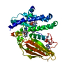
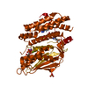
 Basic information
 Components
 Keywords
 Function and homology information
 X-RAY DIFFRACTION /
  Authors
 United States, 1items
  Citation
 Structure visualization
 Downloads & links
 Other downloads


 PDBj
PDBj

 Assembly
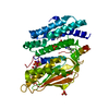


 Mass: 427.201 Da / Num. of mol.: 1 / Source method: obtained synthetically / Formula: C10H15N5O10P2 / Comment: ADP, energy-carrying molecule*YM
 Mass: 24.305 Da / Num. of mol.: 1 / Source method: obtained synthetically / Formula: Mg
 Mass: 39.098 Da / Num. of mol.: 1 / Source method: obtained synthetically / Formula: K
 Mass: 96.063 Da / Num. of mol.: 3 / Source method: obtained synthetically / Formula: SO4
 Mass: 176.175 Da / Num. of mol.: 3 / Source method: obtained synthetically / Formula: C8H8N4O / Feature type: SUBJECT OF INVESTIGATION
 Sample preparation
 Processing