 Movie
Movie Controller

[English] 日本語
 Yorodumi
- PDB-8efr: CryoEM of the soluble OPA1 interfaces with GDP-AlFx bound from th... -
+ Open data

- Basic information
| Entry | Database: PDB / ID: 8efr | ||||||
|---|---|---|---|---|---|---|---|
| Title | CryoEM of the soluble OPA1 interfaces with GDP-AlFx bound from the helical assembly on a lipid membrane | ||||||
 Components Components | Dynamin-like 120 kDa protein, form S1 | ||||||
 Keywords Keywords | LIPID BINDING PROTEIN / GTPase / Dynamin-family protein / mitochondrial fusion protein / mitochondria / Optic Atrophy | ||||||
| Function / homology |  Function and homology information Function and homology informationRegulation of Apoptosis / mitochondrial inner membrane fusion / membrane tubulation / membrane bending activity / GTPase-dependent fusogenic activity / inner mitochondrial membrane organization / dynamin GTPase / cristae formation / peroxisome fission / phosphatidic acid binding ...Regulation of Apoptosis / mitochondrial inner membrane fusion / membrane tubulation / membrane bending activity / GTPase-dependent fusogenic activity / inner mitochondrial membrane organization / dynamin GTPase / cristae formation / peroxisome fission / phosphatidic acid binding / mitochondrial fission / cardiolipin binding / GTP metabolic process / mitochondrial fusion / mitochondrial crista / negative regulation of release of cytochrome c from mitochondria / axonal transport of mitochondrion / positive regulation of interleukin-17 production / protein complex oligomerization / positive regulation of T-helper 17 cell lineage commitment / negative regulation of endoplasmic reticulum stress-induced intrinsic apoptotic signaling pathway / axon cytoplasm / visual perception / Mitochondrial protein degradation / neural tube closure / mitochondrion organization / mitochondrial intermembrane space / mitochondrial membrane / cellular senescence / microtubule binding / microtubule / mitochondrial outer membrane / mitochondrial inner membrane / GTPase activity / apoptotic process / dendrite / negative regulation of apoptotic process / GTP binding / magnesium ion binding / mitochondrion / nucleoplasm / membrane / cytoplasm / cytosol Similarity search - Function | ||||||
| Biological species |  Homo sapiens (human) Homo sapiens (human) | ||||||
| Method | ELECTRON MICROSCOPY / helical reconstruction / cryo EM / Resolution: 5.48 Å | ||||||
 Authors Authors | Nyenhuis, S.B. / Wu, X. / Stanton, A.E. / Strub, M.P. / Yim, Y.I. / Canagarajah, B. / Hinshaw, J.E. | ||||||
| Funding support |  United States, 1items United States, 1items
| ||||||
 Citation Citation | Journal: Nature / Year: 2023 Title: OPA1 helical structures give perspective to mitochondrial dysfunction. Authors: Sarah B Nyenhuis / Xufeng Wu / Marie-Paule Strub / Yang-In Yim / Abigail E Stanton / Valentina Baena / Zulfeqhar A Syed / Bertram Canagarajah / John A Hammer / Jenny E Hinshaw / Abstract: Dominant optic atrophy is one of the leading causes of childhood blindness. Around 60-80% of cases are caused by mutations of the gene that encodes optic atrophy protein 1 (OPA1), a protein that has ...Dominant optic atrophy is one of the leading causes of childhood blindness. Around 60-80% of cases are caused by mutations of the gene that encodes optic atrophy protein 1 (OPA1), a protein that has a key role in inner mitochondrial membrane fusion and remodelling of cristae and is crucial for the dynamic organization and regulation of mitochondria. Mutations in OPA1 result in the dysregulation of the GTPase-mediated fusion process of the mitochondrial inner and outer membranes. Here we used cryo-electron microscopy methods to solve helical structures of OPA1 assembled on lipid membrane tubes, in the presence and absence of nucleotide. These helical assemblies organize into densely packed protein rungs with minimal inter-rung connectivity, and exhibit nucleotide-dependent dimerization of the GTPase domains-a hallmark of the dynamin superfamily of proteins. OPA1 also contains several unique secondary structures in the paddle domain that strengthen its membrane association, including membrane-inserting helices. The structural features identified in this study shed light on the effects of pathogenic point mutations on protein folding, inter-protein assembly and membrane interactions. Furthermore, mutations that disrupt the assembly interfaces and membrane binding of OPA1 cause mitochondrial fragmentation in cell-based assays, providing evidence of the biological relevance of these interactions. | ||||||
| History |
|
- Structure visualization
| Structure viewer | Molecule: MolmilJmol/JSmol |
|---|
- Downloads & links
-Download
| PDBx/mmCIF format | 8efr.cif.gz | 4.5 MB | Display | PDBx/mmCIF format |
|---|---|---|---|---|
| PDB format | pdb8efr.ent.gz | Display | PDB format | |
| PDBx/mmJSON format | 8efr.json.gz | Tree view | PDBx/mmJSON format | |
| Others |  Other downloads Other downloads |
-Validation report
| Arichive directory | https://data.pdbj.org/pub/pdb/validation_reports/ef/8efrftp://data.pdbj.org/pub/pdb/validation_reports/ef/8efr | HTTPS FTP |
|---|
-Related structure data
| Related structure data |  28063MC 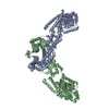 8eewC  8ef7C 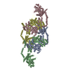 8effC  8efsC  8eftC M: map data used to model this data C: citing same article ( |
|---|---|
| Similar structure data |


-Links
 PDBj
PDBj


- Assembly
| Deposited unit | 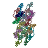
|
|---|---|
| 1 |
|
-Components
| #1: Protein | Mass: 89262.516 Da / Num. of mol.: 18 Source method: isolated from a genetically manipulated source Source: (gene. exp.) Homo sapiens (human) / Gene: OPA1, KIAA0567 / Plasmid: pET28a / Production host:  #2: Chemical | ChemComp-GDP /   Type: RNA linking / Mass: 443.201 Da / Num. of mol.: 18 / Source method: obtained synthetically / Formula: C10H15N5O11P2 / Feature type: SUBJECT OF INVESTIGATION / Comment: GDP, energy-carrying molecule*YM Type: RNA linking / Mass: 443.201 Da / Num. of mol.: 18 / Source method: obtained synthetically / Formula: C10H15N5O11P2 / Feature type: SUBJECT OF INVESTIGATION / Comment: GDP, energy-carrying molecule*YM#3: Chemical | ChemComp-ALF /   Mass: 102.975 Da / Num. of mol.: 18 / Source method: obtained synthetically / Formula: AlF4 / Feature type: SUBJECT OF INVESTIGATION Mass: 102.975 Da / Num. of mol.: 18 / Source method: obtained synthetically / Formula: AlF4 / Feature type: SUBJECT OF INVESTIGATION#4: Chemical | ChemComp-MG /   Mass: 24.305 Da / Num. of mol.: 18 / Source method: obtained synthetically / Formula: Mg / Feature type: SUBJECT OF INVESTIGATION Mass: 24.305 Da / Num. of mol.: 18 / Source method: obtained synthetically / Formula: Mg / Feature type: SUBJECT OF INVESTIGATION#5: Chemical | ChemComp-K /   Mass: 39.098 Da / Num. of mol.: 18 / Source method: obtained synthetically / Formula: K / Feature type: SUBJECT OF INVESTIGATION Mass: 39.098 Da / Num. of mol.: 18 / Source method: obtained synthetically / Formula: K / Feature type: SUBJECT OF INVESTIGATIONHas ligand of interest | Y | Has protein modification | Y | |
|---|
-Experimental details
-Experiment
| Experiment | Method: ELECTRON MICROSCOPY |
|---|---|
| EM experiment | Aggregation state: HELICAL ARRAY / 3D reconstruction method: helical reconstruction |
- Sample preparation
| Component | Name: cryoEM of the soluble OPA1 interfaces with GDP-AlFx bound from the helical assembly on a lipid membrane Type: COMPLEX / Entity ID: #1 / Source: RECOMBINANT |
|---|---|
| Source (natural) | Organism: Homo sapiens (human) |
| Source (recombinant) | Organism: |
| Buffer solution | pH: 7.2 |
| Specimen | Embedding applied: NO / Shadowing applied: NO / Staining applied: NO / Vitrification applied: YES |
| Vitrification | Cryogen name: ETHANE |
- Electron microscopy imaging
| Experimental equipment |  Model: Titan Krios / Image courtesy: FEI Company |
|---|---|
| Microscopy | Model: FEI TITAN KRIOS |
| Electron gun | Electron source:  FIELD EMISSION GUN / Accelerating voltage: 300 kV / Illumination mode: FLOOD BEAM FIELD EMISSION GUN / Accelerating voltage: 300 kV / Illumination mode: FLOOD BEAM |
| Electron lens | Mode: BRIGHT FIELD / Nominal defocus max: 2400 nm / Nominal defocus min: 300 nm |
| Image recording | Electron dose: 60 e/Å2 / Film or detector model: GATAN K3 BIOQUANTUM (6k x 4k) |
- Processing
| EM software |
| |||||||||
|---|---|---|---|---|---|---|---|---|---|---|
| CTF correction | Type: PHASE FLIPPING AND AMPLITUDE CORRECTION | |||||||||
| Helical symmerty | Angular rotation/subunit: 37.439 ° / Axial rise/subunit: 12.933 Å / Axial symmetry: C1 | |||||||||
| 3D reconstruction | Resolution: 5.48 Å / Resolution method: FSC 0.143 CUT-OFF / Num. of particles: 598503 / Symmetry type: HELICAL | |||||||||
| Atomic model building | Protocol: FLEXIBLE FIT / Space: REAL |
