National Institutes of Health/Eunice Kennedy Shriver National Institute of Child Health & Human Development (NIH/NICHD)
R01GM125885
米国
National Institutes of Health/Eunice Kennedy Shriver National Institute of Child Health & Human Development (NIH/NICHD)
P30CA021765
米国
引用
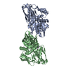
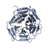
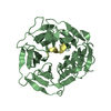
ジャーナル: Mol Cell / 年: 2023 タイトル: E3 ligase autoinhibition by C-degron mimicry maintains C-degron substrate fidelity. 著者: Daniel C Scott / Moeko T King / Kheewoong Baek / Clifford T Gee / Ravi Kalathur / Jerry Li / Nicholas Purser / Amanda Nourse / Sergio C Chai / Sivaraja Vaithiyalingam / Taosheng Chen / ...著者: Daniel C Scott / Moeko T King / Kheewoong Baek / Clifford T Gee / Ravi Kalathur / Jerry Li / Nicholas Purser / Amanda Nourse / Sergio C Chai / Sivaraja Vaithiyalingam / Taosheng Chen / Richard E Lee / Stephen J Elledge / Gary Kleiger / Brenda A Schulman / 要旨: E3 ligase recruitment of proteins containing terminal destabilizing motifs (degrons) is emerging as a major form of regulation. How those E3s discriminate bona fide substrates from other proteins ...E3 ligase recruitment of proteins containing terminal destabilizing motifs (degrons) is emerging as a major form of regulation. How those E3s discriminate bona fide substrates from other proteins with terminal degron-like sequences remains unclear. Here, we report that human KLHDC2, a CRL2 substrate receptor targeting C-terminal Gly-Gly degrons, is regulated through interconversion between two assemblies. In the self-inactivated homotetramer, KLHDC2's C-terminal Gly-Ser motif mimics a degron and engages the substrate-binding domain of another protomer. True substrates capture the monomeric CRL2, driving E3 activation by neddylation and subsequent substrate ubiquitylation. Non-substrates such as NEDD8 bind KLHDC2 with high affinity, but its slow on rate prevents productive association with CRL2. Without substrate, neddylated CRL2 assemblies are deactivated via distinct mechanisms: the monomer by deneddylation and the tetramer by auto-ubiquitylation. Thus, substrate specificity is amplified by KLHDC2 self-assembly acting like a molecular timer, where only bona fide substrates may bind before E3 ligase inactivation.
 ムービー
ムービー コントローラー


 データを開く
 基本情報
基本情報 要素
要素 キーワード
キーワード 機能・相同性情報
 Homo sapiens (ヒト)
 X線回折 /
  データ登録者
 米国, 2件
  引用
引用
 構造の表示
構造の表示 ダウンロードとリンク
 その他のダウンロード


 PDBj
PDBj 集合体
集合体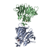

 分子量: 18.015 Da / 分子数: 587 / 由来タイプ: 天然 / 式: H2O
 試料調製
試料調製 解析
解析