 Movie
Movie Controller

[English] 日本語
 Yorodumi
- PDB-8e4f: Crystal structure of dihydrofolate reductase (DHFR) from the fila... -
+ Open data

- Basic information
| Entry | Database: PDB / ID: 8e4f | ||||||
|---|---|---|---|---|---|---|---|
| Title | Crystal structure of dihydrofolate reductase (DHFR) from the filarial nematode W. bancrofti in complex with NADPH and folate | ||||||
 Components Components | Dihydrofolate reductase | ||||||
 Keywords Keywords | OXIDOREDUCTASE / Reductase | ||||||
| Function / homology |  Function and homology information Function and homology informationdihydrofolate metabolic process / dihydrofolate reductase / dihydrofolate reductase activity / folic acid metabolic process / tetrahydrofolate biosynthetic process / one-carbon metabolic process / NADP binding / mitochondrion Similarity search - Function | ||||||
| Biological species |  Wuchereria bancrofti (agent of lymphatic filariasis) Wuchereria bancrofti (agent of lymphatic filariasis) | ||||||
| Method |  X-RAY DIFFRACTION / SYNCHROTRON / MOLECULAR REPLACEMENT / Resolution: 2.47 Å X-RAY DIFFRACTION / SYNCHROTRON / MOLECULAR REPLACEMENT / Resolution: 2.47 Å | ||||||
 Authors Authors | Lange, K. / Frey, K.M. / Goodey, N.M. | ||||||
| Funding support |  United States, 1items United States, 1items
| ||||||
 Citation Citation | Journal: Plos Negl Trop Dis / Year: 2023 Title: Crystal structure of dihydrofolate reductase from the filarial nematode W. bancrofti in complex with NADPH and folate. Authors: Lange, K. / Frey, K.M. / Eck, T. / Janson, C.A. / Gubler, U. / Goodey, N.M. | ||||||
| History |
|
- Structure visualization
| Structure viewer | Molecule: MolmilJmol/JSmol |
|---|
- Downloads & links
-Download
| PDBx/mmCIF format | 8e4f.cif.gz | 57.8 KB | Display | PDBx/mmCIF format |
|---|---|---|---|---|
| PDB format | pdb8e4f.ent.gz | 38.2 KB | Display | PDB format |
| PDBx/mmJSON format | 8e4f.json.gz | Tree view | PDBx/mmJSON format | |
| Others |  Other downloads Other downloads |
-Validation report
| Arichive directory | https://data.pdbj.org/pub/pdb/validation_reports/e4/8e4fftp://data.pdbj.org/pub/pdb/validation_reports/e4/8e4f | HTTPS FTP |
|---|
-Related structure data
| Related structure data | 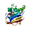 2fzjS S: Starting model for refinement |
|---|---|
| Similar structure data |
-Links
 PDBj
PDBj
- Assembly
| Deposited unit | 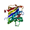
| |||||||||
|---|---|---|---|---|---|---|---|---|---|---|
| 1 |
| |||||||||
| Unit cell |
| |||||||||
| Components on special symmetry positions |
|
-Components
| #1: Protein | Mass: 22381.811 Da / Num. of mol.: 1 Source method: isolated from a genetically manipulated source Source: (gene. exp.) Wuchereria bancrofti (agent of lymphatic filariasis)Gene: WUBG_00817 / Production host:  | ||||
|---|---|---|---|---|---|
| #2: Chemical | ChemComp-NAP /   Mass: 743.405 Da / Num. of mol.: 1 / Source method: obtained synthetically / Formula: C21H28N7O17P3 Mass: 743.405 Da / Num. of mol.: 1 / Source method: obtained synthetically / Formula: C21H28N7O17P3 | ||||
| #3: Chemical | ChemComp-FOL / 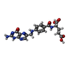  Mass: 441.397 Da / Num. of mol.: 1 / Source method: obtained synthetically / Formula: C19H19N7O6 / Feature type: SUBJECT OF INVESTIGATION Mass: 441.397 Da / Num. of mol.: 1 / Source method: obtained synthetically / Formula: C19H19N7O6 / Feature type: SUBJECT OF INVESTIGATION | ||||
| #4: Chemical |   Mass: 22.990 Da / Num. of mol.: 3 / Source method: obtained synthetically / Formula: Na Mass: 22.990 Da / Num. of mol.: 3 / Source method: obtained synthetically / Formula: Na#5: Water | ChemComp-HOH / |  Mass: 18.015 Da / Num. of mol.: 81 / Source method: isolated from a natural source / Formula: H2O Mass: 18.015 Da / Num. of mol.: 81 / Source method: isolated from a natural source / Formula: H2OHas ligand of interest | Y | |
-Experimental details
-Experiment
| Experiment | Method: X-RAY DIFFRACTION / Number of used crystals: 1 |
|---|
- Sample preparation
| Crystal | Density Matthews: 4.63 Å3/Da / Density % sol: 73.42 % |
|---|---|
| Crystal grow | Temperature: 277.15 K / Method: vapor diffusion, sitting drop Details: 100 mM trisodium citrate pH (5.4-6.4), 25% w/v PEG 4000, and 200 mM ammonium sulfate PH range: 5.4-6.2 |
-Data collection
| Diffraction | Mean temperature: 100 K / Serial crystal experiment: N | ||||||||||||||||||||||||||||||
|---|---|---|---|---|---|---|---|---|---|---|---|---|---|---|---|---|---|---|---|---|---|---|---|---|---|---|---|---|---|---|---|
| Diffraction source | Source: SYNCHROTRON / Site: NSLS-II / Beamline: 17-ID-1 / Wavelength: 1 Å | ||||||||||||||||||||||||||||||
| Detector | Type: DECTRIS EIGER X 9M / Detector: PIXEL / Date: Apr 29, 2019 Details: double crystal Si(111) monochromator with horizontal axis | ||||||||||||||||||||||||||||||
| Radiation | Protocol: SINGLE WAVELENGTH / Monochromatic (M) / Laue (L): M / Scattering type: x-ray | ||||||||||||||||||||||||||||||
| Radiation wavelength | Wavelength: 1 Å / Relative weight: 1 | ||||||||||||||||||||||||||||||
| Reflection | Resolution: 2.47→29.61 Å / Num. obs: 14828 / % possible obs: 99.7 % / Redundancy: 38.5 % / CC1/2: 0.999 / Rmerge(I) obs: 0.164 / Rpim(I) all: 0.027 / Rrim(I) all: 0.167 / Net I/σ(I): 24.8 / Num. measured all: 571238 / Scaling rejects: 2 | ||||||||||||||||||||||||||||||
| Reflection shell | Diffraction-ID: 1
|
- Processing
| Software |
| ||||||||||||||||||||||||||||||||||||||||||
|---|---|---|---|---|---|---|---|---|---|---|---|---|---|---|---|---|---|---|---|---|---|---|---|---|---|---|---|---|---|---|---|---|---|---|---|---|---|---|---|---|---|---|---|
| Refinement | Method to determine structure: MOLECULAR REPLACEMENT Starting model: 2FZJ Resolution: 2.47→29.61 Å / SU ML: 0.25 / Cross valid method: THROUGHOUT / σ(F): 1.34 / Phase error: 22.01 / Stereochemistry target values: ML
| ||||||||||||||||||||||||||||||||||||||||||
| Solvent computation | Shrinkage radii: 0.9 Å / VDW probe radii: 1.11 Å / Solvent model: FLAT BULK SOLVENT MODEL | ||||||||||||||||||||||||||||||||||||||||||
| Displacement parameters | Biso max: 96.3 Å2 / Biso mean: 34.4021 Å2 / Biso min: 15.38 Å2 | ||||||||||||||||||||||||||||||||||||||||||
| Refinement step | Cycle: final / Resolution: 2.47→29.61 Å
| ||||||||||||||||||||||||||||||||||||||||||
| LS refinement shell | Refine-ID: X-RAY DIFFRACTION / Rfactor Rfree error: 0 / Total num. of bins used: 5
|
