 Movie
Movie Controller


+ Open data

- Basic information
| Entry | Database: PDB / ID: 8e1t | ||||||
|---|---|---|---|---|---|---|---|
| Title | Asp1 kinase in complex with ADPNP Mg IP7 | ||||||
 Components Components | Inositol hexakisphosphate and diphosphoinositol-pentakisphosphate kinase | ||||||
 Keywords Keywords | TRANSFERASE / Inositol pyrophosphosphate IPP kinase Fission yeast | ||||||
| Function / homology |  Function and homology information Function and homology informationinositol-1,5-bisdiphosphate-2,3,4,6-tetrakisphosphate 1-diphosphatase activity / regulation of mitotic spindle elongation (spindle phase three) / Synthesis of pyrophosphates in the cytosol / regulation of bipolar cell growth / inositol-1-diphosphate-2,3,4,5,6-pentakisphosphate diphosphatase activity / diphosphoinositol-pentakisphosphate 1-kinase / 5-diphosphoinositol pentakisphosphate 1-kinase activity / inositol hexakisphosphate 1-kinase activity / inositol hexakisphosphate kinase activity / inositol phosphate metabolic process ...inositol-1,5-bisdiphosphate-2,3,4,6-tetrakisphosphate 1-diphosphatase activity / regulation of mitotic spindle elongation (spindle phase three) / Synthesis of pyrophosphates in the cytosol / regulation of bipolar cell growth / inositol-1-diphosphate-2,3,4,5,6-pentakisphosphate diphosphatase activity / diphosphoinositol-pentakisphosphate 1-kinase / 5-diphosphoinositol pentakisphosphate 1-kinase activity / inositol hexakisphosphate 1-kinase activity / inositol hexakisphosphate kinase activity / inositol phosphate metabolic process / inositol phosphate biosynthetic process / inositol phosphate catabolic process / signaling / intracellular phosphate ion homeostasis / regulation of microtubule cytoskeleton organization / 2 iron, 2 sulfur cluster binding / cytoskeleton / ATP hydrolysis activity / ATP binding / nucleus / cytosol Similarity search - Function | ||||||
| Biological species |  | ||||||
| Method |  X-RAY DIFFRACTION / SYNCHROTRON / MOLECULAR REPLACEMENT / Resolution: 1.71 Å X-RAY DIFFRACTION / SYNCHROTRON / MOLECULAR REPLACEMENT / Resolution: 1.71 Å | ||||||
 Authors Authors | Goldgur, Y. / Shuman, S. / Benjamin, B. | ||||||
| Funding support |  United States, 1items United States, 1items
| ||||||
 Citation Citation | Journal: Mbio / Year: 2022 Title: Structures of Fission Yeast Inositol Pyrophosphate Kinase Asp1 in Ligand-Free, Substrate-Bound, and Product-Bound States. Authors: Benjamin, B. / Goldgur, Y. / Jork, N. / Jessen, H.J. / Schwer, B. / Shuman, S. | ||||||
| History |
|
- Structure visualization
| Structure viewer | Molecule: MolmilJmol/JSmol |
|---|
- Downloads & links
-Download
| PDBx/mmCIF format | 8e1t.cif.gz | 164.2 KB | Display | PDBx/mmCIF format |
|---|---|---|---|---|
| PDB format | pdb8e1t.ent.gz | 124 KB | Display | PDB format |
| PDBx/mmJSON format | 8e1t.json.gz | Tree view | PDBx/mmJSON format | |
| Others |  Other downloads Other downloads |
-Validation report
| Arichive directory | https://data.pdbj.org/pub/pdb/validation_reports/e1/8e1tftp://data.pdbj.org/pub/pdb/validation_reports/e1/8e1t | HTTPS FTP |
|---|
-Related structure data
| Related structure data | 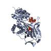 8e1hSC 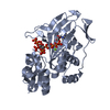 8e1iC 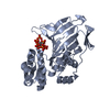 8e1jC 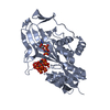 8e1sC 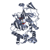 8e1vC S: Starting model for refinement C: citing same article ( |
|---|---|
| Similar structure data |
-Links
 PDBj
PDBj


- Assembly
| Deposited unit | 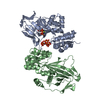
| ||||||||
|---|---|---|---|---|---|---|---|---|---|
| 1 | 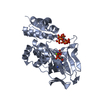
| ||||||||
| 2 | 
| ||||||||
| Unit cell |
|
-Components
| #1: Protein | Mass: 38357.867 Da / Num. of mol.: 2 Source method: isolated from a genetically manipulated source Source: (gene. exp.) Strain: 972 / ATCC 24843 / Gene: asp1, vip1, SPCC1672.06c / Production host:  References: UniProt: O74429, diphosphoinositol-pentakisphosphate 1-kinase #2: Chemical | ChemComp-ANP / |   Mass: 506.196 Da / Num. of mol.: 1 / Source method: obtained synthetically / Formula: C10H17N6O12P3 / Feature type: SUBJECT OF INVESTIGATION / Comment: AMP-PNP, energy-carrying molecule analogue*YM Mass: 506.196 Da / Num. of mol.: 1 / Source method: obtained synthetically / Formula: C10H17N6O12P3 / Feature type: SUBJECT OF INVESTIGATION / Comment: AMP-PNP, energy-carrying molecule analogue*YM#3: Chemical | ChemComp-I7P / ( |   Mass: 740.015 Da / Num. of mol.: 1 / Source method: obtained synthetically / Formula: C6H19O27P7 / Feature type: SUBJECT OF INVESTIGATION Mass: 740.015 Da / Num. of mol.: 1 / Source method: obtained synthetically / Formula: C6H19O27P7 / Feature type: SUBJECT OF INVESTIGATION#4: Chemical | ChemComp-MG / |   Mass: 24.305 Da / Num. of mol.: 1 / Source method: obtained synthetically / Formula: Mg / Feature type: SUBJECT OF INVESTIGATION Mass: 24.305 Da / Num. of mol.: 1 / Source method: obtained synthetically / Formula: Mg / Feature type: SUBJECT OF INVESTIGATION#5: Water | ChemComp-HOH / |  Mass: 18.015 Da / Num. of mol.: 661 / Source method: isolated from a natural source / Formula: H2O Mass: 18.015 Da / Num. of mol.: 661 / Source method: isolated from a natural source / Formula: H2OHas ligand of interest | Y | |
|---|
-Experimental details
-Experiment
| Experiment | Method: X-RAY DIFFRACTION / Number of used crystals: 1 |
|---|
- Sample preparation
| Crystal | Density Matthews: 2.33 Å3/Da / Density % sol: 47.13 % |
|---|---|
| Crystal grow | Temperature: 293 K / Method: vapor diffusion, sitting drop Details: 0.1 M bis-tris propane (pH 8.1), 0.15 M NaF, and 20% PEG 3,350 Temp details: ambient |
-Data collection
| Diffraction | Mean temperature: 100 K / Serial crystal experiment: N | |||||||||||||||||||||||||||||||||||||||||||||||||||||||||||||||||||||||||||||||||||||||||||||||||||||||||||||||||||||||||||||||||||||||||||||||||||||||||||||||||||||||||||||||||||||||||||||
|---|---|---|---|---|---|---|---|---|---|---|---|---|---|---|---|---|---|---|---|---|---|---|---|---|---|---|---|---|---|---|---|---|---|---|---|---|---|---|---|---|---|---|---|---|---|---|---|---|---|---|---|---|---|---|---|---|---|---|---|---|---|---|---|---|---|---|---|---|---|---|---|---|---|---|---|---|---|---|---|---|---|---|---|---|---|---|---|---|---|---|---|---|---|---|---|---|---|---|---|---|---|---|---|---|---|---|---|---|---|---|---|---|---|---|---|---|---|---|---|---|---|---|---|---|---|---|---|---|---|---|---|---|---|---|---|---|---|---|---|---|---|---|---|---|---|---|---|---|---|---|---|---|---|---|---|---|---|---|---|---|---|---|---|---|---|---|---|---|---|---|---|---|---|---|---|---|---|---|---|---|---|---|---|---|---|---|---|---|---|---|
| Diffraction source | Source: SYNCHROTRON / Site: APS / Beamline: 24-ID-E / Wavelength: 0.9792 Å | |||||||||||||||||||||||||||||||||||||||||||||||||||||||||||||||||||||||||||||||||||||||||||||||||||||||||||||||||||||||||||||||||||||||||||||||||||||||||||||||||||||||||||||||||||||||||||||
| Detector | Type: DECTRIS EIGER X 9M / Detector: PIXEL / Date: Apr 26, 2022 | |||||||||||||||||||||||||||||||||||||||||||||||||||||||||||||||||||||||||||||||||||||||||||||||||||||||||||||||||||||||||||||||||||||||||||||||||||||||||||||||||||||||||||||||||||||||||||||
| Radiation | Protocol: SINGLE WAVELENGTH / Monochromatic (M) / Laue (L): M / Scattering type: x-ray | |||||||||||||||||||||||||||||||||||||||||||||||||||||||||||||||||||||||||||||||||||||||||||||||||||||||||||||||||||||||||||||||||||||||||||||||||||||||||||||||||||||||||||||||||||||||||||||
| Radiation wavelength | Wavelength: 0.9792 Å / Relative weight: 1 | |||||||||||||||||||||||||||||||||||||||||||||||||||||||||||||||||||||||||||||||||||||||||||||||||||||||||||||||||||||||||||||||||||||||||||||||||||||||||||||||||||||||||||||||||||||||||||||
| Reflection | Resolution: 1.71→50 Å / Num. obs: 138213 / % possible obs: 95.9 % / Redundancy: 3.9 % / Biso Wilson estimate: 20.27 Å2 / Rmerge(I) obs: 0.056 / Rpim(I) all: 0.033 / Rrim(I) all: 0.065 / Χ2: 1.471 / Net I/σ(I): 11.8 | |||||||||||||||||||||||||||||||||||||||||||||||||||||||||||||||||||||||||||||||||||||||||||||||||||||||||||||||||||||||||||||||||||||||||||||||||||||||||||||||||||||||||||||||||||||||||||||
| Reflection shell | Diffraction-ID: 1
|
- Processing
| Software |
| ||||||||||||||||||||||||||||||||||||||||||||||||||||||||||||||||||||||||||||||||||||||||||||||||||||||||||||||||||||||||||||||||||||||||||||||||||||||||||||||||||||||||||||||||||||||||||||||||||||
|---|---|---|---|---|---|---|---|---|---|---|---|---|---|---|---|---|---|---|---|---|---|---|---|---|---|---|---|---|---|---|---|---|---|---|---|---|---|---|---|---|---|---|---|---|---|---|---|---|---|---|---|---|---|---|---|---|---|---|---|---|---|---|---|---|---|---|---|---|---|---|---|---|---|---|---|---|---|---|---|---|---|---|---|---|---|---|---|---|---|---|---|---|---|---|---|---|---|---|---|---|---|---|---|---|---|---|---|---|---|---|---|---|---|---|---|---|---|---|---|---|---|---|---|---|---|---|---|---|---|---|---|---|---|---|---|---|---|---|---|---|---|---|---|---|---|---|---|---|---|---|---|---|---|---|---|---|---|---|---|---|---|---|---|---|---|---|---|---|---|---|---|---|---|---|---|---|---|---|---|---|---|---|---|---|---|---|---|---|---|---|---|---|---|---|---|---|---|
| Refinement | Method to determine structure: MOLECULAR REPLACEMENT Starting model: 8E1H Resolution: 1.71→47.44 Å / SU ML: 0.17 / Cross valid method: THROUGHOUT / σ(F): 1.34 / Phase error: 21.72 / Stereochemistry target values: ML
| ||||||||||||||||||||||||||||||||||||||||||||||||||||||||||||||||||||||||||||||||||||||||||||||||||||||||||||||||||||||||||||||||||||||||||||||||||||||||||||||||||||||||||||||||||||||||||||||||||||
| Solvent computation | Shrinkage radii: 0.9 Å / VDW probe radii: 1.11 Å / Solvent model: FLAT BULK SOLVENT MODEL | ||||||||||||||||||||||||||||||||||||||||||||||||||||||||||||||||||||||||||||||||||||||||||||||||||||||||||||||||||||||||||||||||||||||||||||||||||||||||||||||||||||||||||||||||||||||||||||||||||||
| Displacement parameters | Biso max: 90.91 Å2 / Biso mean: 25.6469 Å2 / Biso min: 9.42 Å2 | ||||||||||||||||||||||||||||||||||||||||||||||||||||||||||||||||||||||||||||||||||||||||||||||||||||||||||||||||||||||||||||||||||||||||||||||||||||||||||||||||||||||||||||||||||||||||||||||||||||
| Refinement step | Cycle: final / Resolution: 1.71→47.44 Å
| ||||||||||||||||||||||||||||||||||||||||||||||||||||||||||||||||||||||||||||||||||||||||||||||||||||||||||||||||||||||||||||||||||||||||||||||||||||||||||||||||||||||||||||||||||||||||||||||||||||
| LS refinement shell | Refine-ID: X-RAY DIFFRACTION / Rfactor Rfree error: 0 / Total num. of bins used: 27
|
