 Movie
Movie Controller


+ Open data

- Basic information
| Entry | Database: PDB / ID: 8e0r | |||||||||
|---|---|---|---|---|---|---|---|---|---|---|
| Title | Crystal structure of mouse APCDD1 in P21 space group | |||||||||
 Components Components | Protein APCDD1 | |||||||||
 Keywords Keywords | SIGNALING PROTEIN / cell signaling protein / beta barrel / lipid binding protein | |||||||||
| Function / homology |  Function and homology information Function and homology informationregulation of odontogenesis of dentin-containing tooth / astrocyte cell migration / Wnt-protein binding / negative regulation of Wnt signaling pathway / hair follicle development / Wnt signaling pathway / identical protein binding / plasma membrane Similarity search - Function | |||||||||
| Biological species |  | |||||||||
| Method |  X-RAY DIFFRACTION / SYNCHROTRON / MOLECULAR REPLACEMENT / molecular replacement / Resolution: 1.95 Å X-RAY DIFFRACTION / SYNCHROTRON / MOLECULAR REPLACEMENT / molecular replacement / Resolution: 1.95 Å | |||||||||
 Authors Authors | Hsieh, F.L. / Chang, T.H. / Gabelli, S.B. / Nathans, J. | |||||||||
| Funding support |  United States, 2items United States, 2items
| |||||||||
 Citation Citation | Journal: Proc.Natl.Acad.Sci.USA / Year: 2023 Title: Structure of WNT inhibitor adenomatosis polyposis coli down-regulated 1 (APCDD1), a cell-surface lipid-binding protein. Authors: Hsieh, F.L. / Chang, T.H. / Gabelli, S.B. / Nathans, J. | |||||||||
| History |
|
- Structure visualization
| Structure viewer | Molecule: MolmilJmol/JSmol |
|---|
- Downloads & links
-Download
| PDBx/mmCIF format | 8e0r.cif.gz | 345.1 KB | Display | PDBx/mmCIF format |
|---|---|---|---|---|
| PDB format | pdb8e0r.ent.gz | Display | PDB format | |
| PDBx/mmJSON format | 8e0r.json.gz | Tree view | PDBx/mmJSON format | |
| Others |  Other downloads Other downloads |
-Validation report
| Arichive directory | https://data.pdbj.org/pub/pdb/validation_reports/e0/8e0rftp://data.pdbj.org/pub/pdb/validation_reports/e0/8e0r | HTTPS FTP |
|---|
-Related structure data
| Related structure data | 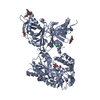 8e0pSC  8e0wC S: Starting model for refinement C: citing same article ( |
|---|---|
| Similar structure data |
-Links
 PDBj
PDBj- Assembly
| Deposited unit | 
| ||||||||||||||||||
|---|---|---|---|---|---|---|---|---|---|---|---|---|---|---|---|---|---|---|---|
| 1 | 
| ||||||||||||||||||
| 2 | 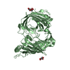
| ||||||||||||||||||
| Unit cell |
| ||||||||||||||||||
| Noncrystallographic symmetry (NCS) | NCS domain:
NCS domain segments: Component-ID: _ / Ens-ID: 1 / Beg auth comp-ID: SER / Beg label comp-ID: SER / End auth comp-ID: SER / End label comp-ID: SER / Refine code: _ / Auth seq-ID: 50 - 468 / Label seq-ID: 27 - 445
|
-Components
| #1: Protein | Mass: 53771.316 Da / Num. of mol.: 2 / Fragment: extracellular domain (UNP residues 27-482) Source method: isolated from a genetically manipulated source Source: (gene. exp.)  Homo sapiens (human) / References: UniProt: Q3U128 Homo sapiens (human) / References: UniProt: Q3U128#2: Sugar | ChemComp-NAG /   Type: D-saccharide, beta linking / Mass: 221.208 Da / Num. of mol.: 5 / Source method: obtained synthetically / Formula: C8H15NO6 Type: D-saccharide, beta linking / Mass: 221.208 Da / Num. of mol.: 5 / Source method: obtained synthetically / Formula: C8H15NO6#3: Water | ChemComp-HOH / |  Mass: 18.015 Da / Num. of mol.: 374 / Source method: isolated from a natural source / Formula: H2O Mass: 18.015 Da / Num. of mol.: 374 / Source method: isolated from a natural source / Formula: H2OHas ligand of interest | N | Has protein modification | Y | |
|---|
-Experimental details
-Experiment
| Experiment | Method: X-RAY DIFFRACTION / Number of used crystals: 1 |
|---|
- Sample preparation
| Crystal | Density Matthews: 2.12 Å3/Da / Density % sol: 42.02 % |
|---|---|
| Crystal grow | Temperature: 293 K / Method: vapor diffusion / pH: 7 Details: 0.7 M magnesium formate, 0.1 M Bis-Tris propane, pH 7.0 |
-Data collection
| Diffraction | Mean temperature: 100 K / Serial crystal experiment: N |
|---|---|
| Diffraction source | Source: SYNCHROTRON / Site: NSLS-II / Beamline: 17-ID-2 / Wavelength: 0.92009 Å |
| Detector | Type: DECTRIS EIGER X 16M / Detector: PIXEL / Date: Apr 10, 2019 |
| Radiation | Protocol: SINGLE WAVELENGTH / Monochromatic (M) / Laue (L): M / Scattering type: x-ray |
| Radiation wavelength | Wavelength: 0.92009 Å / Relative weight: 1 |
| Reflection | Resolution: 1.947→34.364 Å / Num. obs: 49930 / % possible obs: 91.1 % / Redundancy: 6.3 % / CC1/2: 0.996 / Rmerge(I) obs: 0.135 / Rpim(I) all: 0.057 / Net I/σ(I): 8.5 |
| Reflection shell | Resolution: 1.947→2.12 Å / Rmerge(I) obs: 1.464 / Mean I/σ(I) obs: 1.3 / Num. unique obs: 2486 / CC1/2: 0.56 / Rpim(I) all: 0.58 |
-Phasing
| Phasing | Method: molecular replacement | |||||||||
|---|---|---|---|---|---|---|---|---|---|---|
| Phasing MR |
|
- Processing
| Software |
| |||||||||||||||||||||||||||||||||||||||||||||||||||||||||||||||||||||||||||
|---|---|---|---|---|---|---|---|---|---|---|---|---|---|---|---|---|---|---|---|---|---|---|---|---|---|---|---|---|---|---|---|---|---|---|---|---|---|---|---|---|---|---|---|---|---|---|---|---|---|---|---|---|---|---|---|---|---|---|---|---|---|---|---|---|---|---|---|---|---|---|---|---|---|---|---|---|
| Refinement | Method to determine structure: MOLECULAR REPLACEMENT Starting model: PDB entry 8E0P Resolution: 1.95→34 Å / Cor.coef. Fo:Fc: 0.947 / Cor.coef. Fo:Fc free: 0.926 / SU B: 12.349 / SU ML: 0.168 / Cross valid method: THROUGHOUT / σ(F): 0 / ESU R: 0.282 / ESU R Free: 0.208 / Stereochemistry target values: MAXIMUM LIKELIHOOD Details: U VALUES : TLS ONLY HYDROGENS HAVE BEEN ADDED IN THE RIDING POSITIONS
| |||||||||||||||||||||||||||||||||||||||||||||||||||||||||||||||||||||||||||
| Solvent computation | Ion probe radii: 0.8 Å / Shrinkage radii: 0.8 Å / VDW probe radii: 1.2 Å / Solvent model: MASK | |||||||||||||||||||||||||||||||||||||||||||||||||||||||||||||||||||||||||||
| Displacement parameters | Biso max: 68.1 Å2 / Biso mean: 9.798 Å2 / Biso min: 1.4 Å2
| |||||||||||||||||||||||||||||||||||||||||||||||||||||||||||||||||||||||||||
| Refinement step | Cycle: final / Resolution: 1.95→34 Å
| |||||||||||||||||||||||||||||||||||||||||||||||||||||||||||||||||||||||||||
| Refine LS restraints |
| |||||||||||||||||||||||||||||||||||||||||||||||||||||||||||||||||||||||||||
| Refine LS restraints NCS | Ens-ID: 1 / Number: 12634 / Refine-ID: X-RAY DIFFRACTION / Type: interatomic distance / Rms dev position: 0.12 Å / Weight position: 0.05
| |||||||||||||||||||||||||||||||||||||||||||||||||||||||||||||||||||||||||||
| LS refinement shell | Resolution: 1.95→2 Å / Rfactor Rfree error: 0 / Total num. of bins used: 20
| |||||||||||||||||||||||||||||||||||||||||||||||||||||||||||||||||||||||||||
| Refinement TLS params. | Method: refined / Refine-ID: X-RAY DIFFRACTION
| |||||||||||||||||||||||||||||||||||||||||||||||||||||||||||||||||||||||||||
| Refinement TLS group |
|
