Movie
Movie Controller

[English] 日本語
 Yorodumi
- PDB-8dtb: Focus/local refined map in C1 of signal subtracted RyR1 particles... -
+ Open data

- Basic information
| Entry | Database: PDB / ID: 8dtb | |||||||||
|---|---|---|---|---|---|---|---|---|---|---|
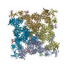
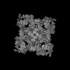
| Title | Focus/local refined map in C1 of signal subtracted RyR1 particles in complex with ImperaCalcin | |||||||||
 Components Components |
| |||||||||
 Keywords Keywords | TOXIN / ryanodine receptor / ion channel / snake toxin / calcin / complex / membrane protein | |||||||||
| Function / homology |  Function and homology information Function and homology informationATP-gated ion channel activity / ryanodine-sensitive calcium-release channel activity / terminal cisterna / ryanodine receptor complex / release of sequestered calcium ion into cytosol by sarcoplasmic reticulum / ossification involved in bone maturation / cellular response to caffeine / skin development / organelle membrane / smooth endoplasmic reticulum ...ATP-gated ion channel activity / ryanodine-sensitive calcium-release channel activity / terminal cisterna / ryanodine receptor complex / release of sequestered calcium ion into cytosol by sarcoplasmic reticulum / ossification involved in bone maturation / cellular response to caffeine / skin development / organelle membrane / smooth endoplasmic reticulum / outflow tract morphogenesis / intracellularly gated calcium channel activity / toxic substance binding / voltage-gated calcium channel activity / calcium channel inhibitor activity / skeletal muscle fiber development / release of sequestered calcium ion into cytosol / muscle contraction / sarcoplasmic reticulum membrane / striated muscle contraction / cellular response to calcium ion / sarcoplasmic reticulum / sarcolemma / intracellular calcium ion homeostasis / Z disc / calcium channel activity / calcium ion transmembrane transport / disordered domain specific binding / toxin activity / protein homotetramerization / transmembrane transporter binding / calmodulin binding / calcium ion binding / extracellular region / ATP binding / membrane / identical protein binding Similarity search - Function | |||||||||
| Biological species |   Pandinus imperator (emperor scorpion) Pandinus imperator (emperor scorpion) | |||||||||
| Method | ELECTRON MICROSCOPY / single particle reconstruction / cryo EM / Resolution: 3.14 Å | |||||||||
 Authors Authors | Haji-Ghassemi, O. / Van Petegm, F. | |||||||||
| Funding support |  Canada, 2items Canada, 2items
| |||||||||
 Citation Citation | Journal: Sci Adv / Year: 2023 Title: Cryo-EM analysis of scorpion toxin binding to Ryanodine Receptors reveals subconductance that is abolished by PKA phosphorylation. Authors: Omid Haji-Ghassemi / Yu Seby Chen / Kellie Woll / Georgina B Gurrola / Carmen R Valdivia / Wenxuan Cai / Songhua Li / Hector H Valdivia / Filip Van Petegem /    Abstract: Calcins are peptides from scorpion venom with the unique ability to cross cell membranes, gaining access to intracellular targets. Ryanodine Receptors (RyR) are intracellular ion channels that ...Calcins are peptides from scorpion venom with the unique ability to cross cell membranes, gaining access to intracellular targets. Ryanodine Receptors (RyR) are intracellular ion channels that control release of Ca from the endoplasmic and sarcoplasmic reticulum. Calcins target RyRs and induce long-lived subconductance states, whereby single-channel currents are decreased. We used cryo-electron microscopy to reveal the binding and structural effects of imperacalcin, showing that it opens the channel pore and causes large asymmetry throughout the cytosolic assembly of the tetrameric RyR. This also creates multiple extended ion conduction pathways beyond the transmembrane region, resulting in subconductance. Phosphorylation of imperacalcin by protein kinase A prevents its binding to RyR through direct steric hindrance, showing how posttranslational modifications made by the host organism can determine the fate of a natural toxin. The structure provides a direct template for developing calcin analogs that result in full channel block, with potential to treat RyR-related disorders. | |||||||||
| History |
|
- Structure visualization
| Structure viewer | Molecule: MolmilJmol/JSmol |
|---|
- Downloads & links
-Download
| PDBx/mmCIF format | 8dtb.cif.gz | 640.3 KB | Display | PDBx/mmCIF format |
|---|---|---|---|---|
| PDB format | pdb8dtb.ent.gz | 347.3 KB | Display | PDB format |
| PDBx/mmJSON format | 8dtb.json.gz | Tree view | PDBx/mmJSON format | |
| Others |  Other downloads Other downloads |
-Validation report
| Arichive directory | https://data.pdbj.org/pub/pdb/validation_reports/dt/8dtbftp://data.pdbj.org/pub/pdb/validation_reports/dt/8dtb | HTTPS FTP |
|---|
-Related structure data
| Related structure data | 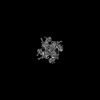 27695MC  8drpC  8dujC  8dveC C: citing same article ( M: map data used to model this data |
|---|---|
| Similar structure data |

-Links
 PDBj
PDBj
- Assembly
| Deposited unit | 
|
|---|---|
| 1 |
|
-Components
| #1: Protein/peptide | Mass: 3776.495 Da / Num. of mol.: 1 / Source method: obtained synthetically / Source: (synth.) Pandinus imperator (emperor scorpion) / References: UniProt: P59868 | ||||||||
|---|---|---|---|---|---|---|---|---|---|
| #2: Protein | Mass: 565908.625 Da / Num. of mol.: 4 / Source method: isolated from a natural source / Source: (natural) #3: Chemical | ChemComp-ATP /   Mass: 507.181 Da / Num. of mol.: 4 / Source method: isolated from a natural source / Formula: C10H16N5O13P3 / Comment: ATP, energy-carrying molecule*YM Mass: 507.181 Da / Num. of mol.: 4 / Source method: isolated from a natural source / Formula: C10H16N5O13P3 / Comment: ATP, energy-carrying molecule*YM#4: Chemical |   Mass: 194.191 Da / Num. of mol.: 3 / Source method: isolated from a natural source / Formula: C8H10N4O2 / Comment: medication*YM Mass: 194.191 Da / Num. of mol.: 3 / Source method: isolated from a natural source / Formula: C8H10N4O2 / Comment: medication*YMHas ligand of interest | N | Has protein modification | Y | |
-Experimental details
-Experiment
| Experiment | Method: ELECTRON MICROSCOPY |
|---|---|
| EM experiment | Aggregation state: PARTICLE / 3D reconstruction method: single particle reconstruction |
- Sample preparation
| Component | Name: RyR1 in complex with ImperaCalcin / Type: COMPLEX / Entity ID: #1-#2 / Source: MULTIPLE SOURCES | ||||||||||||
|---|---|---|---|---|---|---|---|---|---|---|---|---|---|
| Molecular weight | Value: 2.2 MDa / Experimental value: YES | ||||||||||||
| Source (natural) |
| ||||||||||||
| Buffer solution | pH: 7.4 | ||||||||||||
| Specimen | Conc.: 10 mg/ml / Embedding applied: NO / Shadowing applied: NO / Staining applied: NO / Vitrification applied: YES | ||||||||||||
| Specimen support | Grid material: COPPER / Grid mesh size: 300 divisions/in. / Grid type: Quantifoil R2/2 | ||||||||||||
| Vitrification | Cryogen name: ETHANE |
- Electron microscopy imaging
| Experimental equipment |  Model: Titan Krios / Image courtesy: FEI Company |
|---|---|
| Microscopy | Model: FEI TITAN KRIOS |
| Electron gun | Electron source:  FIELD EMISSION GUN / Accelerating voltage: 300 kV / Illumination mode: FLOOD BEAM FIELD EMISSION GUN / Accelerating voltage: 300 kV / Illumination mode: FLOOD BEAM |
| Electron lens | Mode: BRIGHT FIELD / Nominal defocus max: 3000 nm / Nominal defocus min: 1000 nm / Cs: 2.7 mm |
| Image recording | Electron dose: 50 e/Å2 / Detector mode: COUNTING / Film or detector model: FEI FALCON III (4k x 4k) |
| Image scans | Width: 4096 / Height: 4096 |
- Processing
| EM software |
| |||||||||||||||||||||||||||||||||||||||||||||
|---|---|---|---|---|---|---|---|---|---|---|---|---|---|---|---|---|---|---|---|---|---|---|---|---|---|---|---|---|---|---|---|---|---|---|---|---|---|---|---|---|---|---|---|---|---|---|
| CTF correction | Type: PHASE FLIPPING AND AMPLITUDE CORRECTION | |||||||||||||||||||||||||||||||||||||||||||||
| Symmetry | Point symmetry: C1 (asymmetric) | |||||||||||||||||||||||||||||||||||||||||||||
| 3D reconstruction | Resolution: 3.14 Å / Resolution method: FSC 0.143 CUT-OFF / Num. of particles: 307078 / Algorithm: FOURIER SPACE / Num. of class averages: 3 / Symmetry type: POINT | |||||||||||||||||||||||||||||||||||||||||||||
| Atomic model building | Protocol: AB INITIO MODEL | |||||||||||||||||||||||||||||||||||||||||||||
| Atomic model building | PDB-ID: 6M2W Accession code: 6M2W / Source name: PDB / Type: experimental model | |||||||||||||||||||||||||||||||||||||||||||||
| Refinement | Highest resolution: 3.14 Å |