 Movie
Movie Controller


+ Open data

- Basic information
| Entry | Database: PDB / ID: 8dpw | ||||||
|---|---|---|---|---|---|---|---|
| Title | The structure of Interleukin-11 Mutein | ||||||
 Components Components | Interleukin-11 | ||||||
 Keywords Keywords | CYTOKINE / complex / IL-6 family cytokine | ||||||
| Function / homology |  Function and homology information Function and homology informationinterleukin-11 receptor binding / negative regulation of cytokine activity / negative regulation of cytokine-mediated signaling pathway / negative regulation of hormone secretion / megakaryocyte differentiation / interleukin-11-mediated signaling pathway / positive regulation of peptidyl-tyrosine phosphorylation / IL-6-type cytokine receptor ligand interactions / positive regulation of peptidyl-serine phosphorylation / fat cell differentiation ...interleukin-11 receptor binding / negative regulation of cytokine activity / negative regulation of cytokine-mediated signaling pathway / negative regulation of hormone secretion / megakaryocyte differentiation / interleukin-11-mediated signaling pathway / positive regulation of peptidyl-tyrosine phosphorylation / IL-6-type cytokine receptor ligand interactions / positive regulation of peptidyl-serine phosphorylation / fat cell differentiation / B cell differentiation / cytokine activity / growth factor activity / positive regulation of MAPK cascade / positive regulation of cell population proliferation / positive regulation of transcription by RNA polymerase II / : / extracellular region / cytoplasm Similarity search - Function | ||||||
| Biological species |  Homo sapiens (human) Homo sapiens (human) | ||||||
| Method |  X-RAY DIFFRACTION / SYNCHROTRON / MOLECULAR REPLACEMENT / Resolution: 1.8 Å X-RAY DIFFRACTION / SYNCHROTRON / MOLECULAR REPLACEMENT / Resolution: 1.8 Å | ||||||
 Authors Authors | Metcalfe, R.D. / Griffin, M.D.W. | ||||||
| Funding support |  Australia, 1items Australia, 1items
| ||||||
 Citation Citation | Journal: Nat Commun / Year: 2023 Title: Structures of the interleukin 11 signalling complex reveal gp130 dynamics and the inhibitory mechanism of a cytokine variant. Authors: Riley D Metcalfe / Eric Hanssen / Ka Yee Fung / Kaheina Aizel / Clara C Kosasih / Courtney O Zlatic / Larissa Doughty / Craig J Morton / Andrew P Leis / Michael W Parker / Paul R Gooley / ...Authors: Riley D Metcalfe / Eric Hanssen / Ka Yee Fung / Kaheina Aizel / Clara C Kosasih / Courtney O Zlatic / Larissa Doughty / Craig J Morton / Andrew P Leis / Michael W Parker / Paul R Gooley / Tracy L Putoczki / Michael D W Griffin /  Abstract: Interleukin (IL-)11, an IL-6 family cytokine, has pivotal roles in autoimmune diseases, fibrotic complications, and solid cancers. Despite intense therapeutic targeting efforts, structural ...Interleukin (IL-)11, an IL-6 family cytokine, has pivotal roles in autoimmune diseases, fibrotic complications, and solid cancers. Despite intense therapeutic targeting efforts, structural understanding of IL-11 signalling and mechanistic insights into current inhibitors are lacking. Here we present cryo-EM and crystal structures of the human IL-11 signalling complex, including the complex containing the complete extracellular domains of the shared IL-6 family β-receptor, gp130. We show that complex formation requires conformational reorganisation of IL-11 and that the membrane-proximal domains of gp130 are dynamic. We demonstrate that the cytokine mutant, IL-11 Mutein, competitively inhibits signalling in human cell lines. Structural shifts in IL-11 Mutein underlie inhibition by altering cytokine binding interactions at all three receptor-engaging sites and abrogating the final gp130 binding step. Our results reveal the structural basis of IL-11 signalling, define the molecular mechanisms of an inhibitor, and advance understanding of gp130-containing receptor complexes, with potential applications in therapeutic development. | ||||||
| History |
|
- Structure visualization
| Structure viewer | Molecule: MolmilJmol/JSmol |
|---|
- Downloads & links
-Download
| PDBx/mmCIF format | 8dpw.cif.gz | 125.7 KB | Display | PDBx/mmCIF format |
|---|---|---|---|---|
| PDB format | pdb8dpw.ent.gz | 81.9 KB | Display | PDB format |
| PDBx/mmJSON format | 8dpw.json.gz | Tree view | PDBx/mmJSON format | |
| Others |  Other downloads Other downloads |
-Validation report
| Arichive directory | https://data.pdbj.org/pub/pdb/validation_reports/dp/8dpwftp://data.pdbj.org/pub/pdb/validation_reports/dp/8dpw | HTTPS FTP |
|---|
-Related structure data
| Related structure data |  8dpsC  8dptC  8dpuC  8dpvC  4mhlS S: Starting model for refinement C: citing same article ( |
|---|---|
| Similar structure data | |
| Other databases |
|
-Links
 PDBj
PDBj


- Assembly
| Deposited unit | 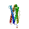
| ||||||||||||
|---|---|---|---|---|---|---|---|---|---|---|---|---|---|
| 1 |
| ||||||||||||
| Unit cell |
|
-Components
| #1: Protein | Mass: 18300.293 Da / Num. of mol.: 1 / Mutation: A58P, M59A, S60I, A61D, G62Y, W147A Source method: isolated from a genetically manipulated source Source: (gene. exp.) Homo sapiens (human) / Gene: IL11 / Production host:  |
|---|---|
| #2: Chemical | ChemComp-SO4 /   Mass: 96.063 Da / Num. of mol.: 1 / Source method: obtained synthetically / Formula: SO4 Mass: 96.063 Da / Num. of mol.: 1 / Source method: obtained synthetically / Formula: SO4 |
| #3: Water | ChemComp-HOH /  Mass: 18.015 Da / Num. of mol.: 71 / Source method: isolated from a natural source / Formula: H2O Mass: 18.015 Da / Num. of mol.: 71 / Source method: isolated from a natural source / Formula: H2O |
| Has ligand of interest | N |
| Has protein modification | N |
-Experimental details
-Experiment
| Experiment | Method: X-RAY DIFFRACTION / Number of used crystals: 1 |
|---|
- Sample preparation
| Crystal | Density Matthews: 1.85 Å3/Da / Density % sol: 33.68 % |
|---|---|
| Crystal grow | Temperature: 293 K / Method: vapor diffusion, sitting drop / pH: 9 Details: 27% PEG 3350, 0.1 M bis-tris propane pH 9, 0.2 M ammonium sulfate, 5 mM praseodymium chloride |
-Data collection
| Diffraction | Mean temperature: 100 K / Serial crystal experiment: N |
|---|---|
| Diffraction source | Source: SYNCHROTRON / Site: Australian Synchrotron / Beamline: MX2 / Wavelength: 0.9537 Å |
| Detector | Type: DECTRIS EIGER X 16M / Detector: PIXEL / Date: Aug 5, 2017 |
| Radiation | Protocol: SINGLE WAVELENGTH / Monochromatic (M) / Laue (L): M / Scattering type: x-ray |
| Radiation wavelength | Wavelength: 0.9537 Å / Relative weight: 1 |
| Reflection | Resolution: 1.8→37.101 Å / Num. obs: 12637 / % possible obs: 100 % / Redundancy: 6.8 % / Biso Wilson estimate: 32.06 Å2 / CC1/2: 0.99 / Rpim(I) all: 0.043 / Rrim(I) all: 0.083 / Rsym value: 0.07 / Net I/σ(I): 11.8 |
| Reflection shell | Resolution: 1.8→1.84 Å / Redundancy: 6.9 % / Mean I/σ(I) obs: 1.4 / Num. unique obs: 761 / CC1/2: 0.603 / Rpim(I) all: 0.719 / Rrim(I) all: 1.374 / Rsym value: 1.167 / % possible all: 100 |
- Processing
| Software |
| ||||||||||||||||||||||||||||||||||||||||
|---|---|---|---|---|---|---|---|---|---|---|---|---|---|---|---|---|---|---|---|---|---|---|---|---|---|---|---|---|---|---|---|---|---|---|---|---|---|---|---|---|---|
| Refinement | Method to determine structure: MOLECULAR REPLACEMENT Starting model: 4MHL Resolution: 1.8→33.6 Å / SU ML: 0.2633 / Cross valid method: FREE R-VALUE / σ(F): 1.41 / Phase error: 28.7198 Stereochemistry target values: GeoStd + Monomer Library + CDL v1.2
| ||||||||||||||||||||||||||||||||||||||||
| Solvent computation | Shrinkage radii: 0.9 Å / VDW probe radii: 1.11 Å / Solvent model: FLAT BULK SOLVENT MODEL | ||||||||||||||||||||||||||||||||||||||||
| Displacement parameters | Biso mean: 46.65 Å2 | ||||||||||||||||||||||||||||||||||||||||
| Refinement step | Cycle: LAST / Resolution: 1.8→33.6 Å
| ||||||||||||||||||||||||||||||||||||||||
| Refine LS restraints |
| ||||||||||||||||||||||||||||||||||||||||
| LS refinement shell |
| ||||||||||||||||||||||||||||||||||||||||
| Refinement TLS params. | Method: refined / Origin x: -6.7465929837 Å / Origin y: 7.63166353347 Å / Origin z: -17.8512695497 Å
| ||||||||||||||||||||||||||||||||||||||||
| Refinement TLS group | Selection details: (chain 'A' and resid 31 through 178) |
