 Movie
Movie Controller

[English] 日本語
 Yorodumi
- PDB-8dk4: Peroxisome proliferator-activated receptor gamma in complex with ... -
+ Open data

- Basic information
| Entry | Database: PDB / ID: 8dk4 | |||||||||
|---|---|---|---|---|---|---|---|---|---|---|
| Title | Peroxisome proliferator-activated receptor gamma in complex with VSP-51-2 | |||||||||
 Components Components |
| |||||||||
 Keywords Keywords | SIGNALING PROTEIN / Peroxisome proliferator-activated receptor gamma / Modulator / VSP-51-2 | |||||||||
| Function / homology |  Function and homology information Function and homology informationRegulation of MITF-M dependent genes involved in metabolism / positive regulation of fatty acid oxidation / cellular respiration / Activation of PPARGC1A (PGC-1alpha) by phosphorylation / prostaglandin receptor activity / : / negative regulation of receptor signaling pathway via STAT / MECP2 regulates transcription factors / negative regulation of vascular endothelial cell proliferation / negative regulation of extracellular matrix assembly ...Regulation of MITF-M dependent genes involved in metabolism / positive regulation of fatty acid oxidation / cellular respiration / Activation of PPARGC1A (PGC-1alpha) by phosphorylation / prostaglandin receptor activity / : / negative regulation of receptor signaling pathway via STAT / MECP2 regulates transcription factors / negative regulation of vascular endothelial cell proliferation / negative regulation of extracellular matrix assembly / negative regulation of connective tissue replacement involved in inflammatory response wound healing / positive regulation of cholesterol transport / negative regulation of cellular response to transforming growth factor beta stimulus / arachidonate binding / positive regulation of adiponectin secretion / negative regulation of cardiac muscle hypertrophy in response to stress / DNA binding domain binding / lipoprotein transport / positive regulation of vascular associated smooth muscle cell apoptotic process / temperature homeostasis / WW domain binding / response to starvation / positive regulation of fatty acid metabolic process / STAT family protein binding / response to lipid / lncRNA binding / response to muscle activity / negative regulation of type II interferon-mediated signaling pathway / LBD domain binding / negative regulation of cholesterol storage / intracellular glucose homeostasis / negative regulation of SMAD protein signal transduction / fatty acid oxidation / response to dietary excess / lipid homeostasis / E-box binding / alpha-actinin binding / R-SMAD binding / negative regulation of vascular associated smooth muscle cell proliferation / monocyte differentiation / negative regulation of blood vessel endothelial cell migration / white fat cell differentiation / cellular response to low-density lipoprotein particle stimulus / negative regulation of macrophage derived foam cell differentiation / negative regulation of lipid storage / negative regulation of BMP signaling pathway / positive regulation of cholesterol efflux / cell fate commitment / adipose tissue development / negative regulation of osteoblast differentiation / negative regulation of mitochondrial fission / positive regulation of fat cell differentiation / FOXO-mediated transcription of oxidative stress, metabolic and neuronal genes / BMP signaling pathway / long-chain fatty acid transport / brown fat cell differentiation / nuclear retinoid X receptor binding / Transcriptional regulation of brown and beige adipocyte differentiation by EBF2 / retinoic acid receptor signaling pathway / energy homeostasis / cell maturation / digestion / negative regulation of MAPK cascade / intracellular receptor signaling pathway / hormone-mediated signaling pathway / positive regulation of adipose tissue development / peroxisome proliferator activated receptor signaling pathway / response to nutrient / positive regulation of gluconeogenesis / epithelial cell differentiation / regulation of cellular response to insulin stimulus / peptide binding / SUMOylation of transcription cofactors / RNA splicing / negative regulation of miRNA transcription / negative regulation of angiogenesis / placenta development / nuclear receptor binding / Regulation of PTEN gene transcription / positive regulation of apoptotic signaling pathway / gluconeogenesis / transcription coregulator binding / respiratory electron transport chain / transcription coregulator activity / mitochondrion organization / transcription initiation at RNA polymerase II promoter / SUMOylation of intracellular receptors / negative regulation of smooth muscle cell proliferation / circadian regulation of gene expression / mRNA transcription by RNA polymerase II / Heme signaling / negative regulation of transforming growth factor beta receptor signaling pathway / fatty acid metabolic process / Transcriptional activation of mitochondrial biogenesis / PPARA activates gene expression / regulation of circadian rhythm / PML body / chromatin DNA binding / Nuclear Receptor transcription pathway / Transcriptional regulation of white adipocyte differentiation Similarity search - Function | |||||||||
| Biological species |  Homo sapiens (human) Homo sapiens (human) | |||||||||
| Method |  X-RAY DIFFRACTION / SYNCHROTRON / MOLECULAR REPLACEMENT / Resolution: 2.6 Å X-RAY DIFFRACTION / SYNCHROTRON / MOLECULAR REPLACEMENT / Resolution: 2.6 Å | |||||||||
 Authors Authors | Ma, L. / Zhou, X.E. / Suino-Powell, K. / Hou, N. / Zhou, Z. / Luo, J. / Xu, H.E. / Yi, W. | |||||||||
| Funding support |  China, 2items China, 2items
| |||||||||
 Citation Citation | Journal: To Be Published Title: Identification of VSP-51-2 as the Novel and Safe PPAR gamma Modulator: Structure-Based Design, Biological Validation and Crystal Analysis Authors: Ma, L. / Zhou, X.E. / Suino-Powell, K. / Hou, N. / Zhou, Z. / Luo, J. / Xu, H.E. / Yi, W. | |||||||||
| History |
|
- Structure visualization
| Structure viewer | Molecule: MolmilJmol/JSmol |
|---|
- Downloads & links
-Download
| PDBx/mmCIF format | 8dk4.cif.gz | 72 KB | Display | PDBx/mmCIF format |
|---|---|---|---|---|
| PDB format | pdb8dk4.ent.gz | 51.2 KB | Display | PDB format |
| PDBx/mmJSON format | 8dk4.json.gz | Tree view | PDBx/mmJSON format | |
| Others |  Other downloads Other downloads |
-Validation report
| Summary document | 8dk4_validation.pdf.gz | 748.4 KB | Display | wwPDB validaton report |
|---|---|---|---|---|
| Full document | 8dk4_full_validation.pdf.gz | 750.5 KB | Display | |
| Data in XML | 8dk4_validation.xml.gz | 12.2 KB | Display | |
| Data in CIF | 8dk4_validation.cif.gz | 15.8 KB | Display | |
| Arichive directory | https://data.pdbj.org/pub/pdb/validation_reports/dk/8dk4ftp://data.pdbj.org/pub/pdb/validation_reports/dk/8dk4 | HTTPS FTP |
-Related structure data
| Related structure data |  1fm6S S: Starting model for refinement |
|---|---|
| Similar structure data |
-Links
 PDBj
PDBj


- Assembly
| Deposited unit | 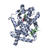
| ||||||||
|---|---|---|---|---|---|---|---|---|---|
| 1 |
| ||||||||
| Unit cell |
|
-Components
| #1: Protein | Mass: 31094.135 Da / Num. of mol.: 1 / Fragment: ligand binding domain (UNP residues 234-505) Source method: isolated from a genetically manipulated source Source: (gene. exp.) Homo sapiens (human) / Gene: PPARG, NR1C3 / Production host:  |
|---|---|
| #2: Protein/peptide | Mass: 1594.932 Da / Num. of mol.: 1 / Fragment: UNP residues 138-152 Source method: isolated from a genetically manipulated source Source: (gene. exp.) Homo sapiens (human) / Gene: PPARGC1A, LEM6, PGC1, PGC1A, PPARGC1 / Production host: |
| #3: Chemical | ChemComp-SJ9 / 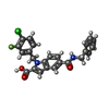  Mass: 436.863 Da / Num. of mol.: 1 / Source method: obtained synthetically / Formula: C24H18ClFN2O3 / Feature type: SUBJECT OF INVESTIGATION Mass: 436.863 Da / Num. of mol.: 1 / Source method: obtained synthetically / Formula: C24H18ClFN2O3 / Feature type: SUBJECT OF INVESTIGATION |
| #4: Water | ChemComp-HOH /  Mass: 18.015 Da / Num. of mol.: 4 / Source method: isolated from a natural source / Formula: H2O Mass: 18.015 Da / Num. of mol.: 4 / Source method: isolated from a natural source / Formula: H2O |
| Has ligand of interest | Y |
-Experimental details
-Experiment
| Experiment | Method: X-RAY DIFFRACTION / Number of used crystals: 1 |
|---|
- Sample preparation
| Crystal | Density Matthews: 2.36 Å3/Da / Density % sol: 47.95 % |
|---|---|
| Crystal grow | Temperature: 293 K / Method: vapor diffusion, sitting drop Details: 0.1 M of trisodium citrate, pH 5.5, 20% w/v PEG3350 |
-Data collection
| Diffraction | Mean temperature: 100 K / Serial crystal experiment: N | ||||||||||||||||||||||||||||||
|---|---|---|---|---|---|---|---|---|---|---|---|---|---|---|---|---|---|---|---|---|---|---|---|---|---|---|---|---|---|---|---|
| Diffraction source | Source: SYNCHROTRON / Site: APS  / Beamline: 21-ID-D / Wavelength: 1 Å / Beamline: 21-ID-D / Wavelength: 1 Å | ||||||||||||||||||||||||||||||
| Detector | Type: DECTRIS EIGER2 S 16M / Detector: PIXEL / Date: Mar 10, 2015 | ||||||||||||||||||||||||||||||
| Radiation | Protocol: SINGLE WAVELENGTH / Monochromatic (M) / Laue (L): M / Scattering type: x-ray | ||||||||||||||||||||||||||||||
| Radiation wavelength | Wavelength: 1 Å / Relative weight: 1 | ||||||||||||||||||||||||||||||
| Reflection | Resolution: 2.6→47.39 Å / Num. obs: 9684 / % possible obs: 100 % / Redundancy: 7.1 % / Biso Wilson estimate: 66.81 Å2 / CC1/2: 0.998 / Rmerge(I) obs: 0.105 / Rpim(I) all: 0.043 / Rrim(I) all: 0.114 / Net I/σ(I): 6.3 / Num. measured all: 69144 / Scaling rejects: 143 | ||||||||||||||||||||||||||||||
| Reflection shell | Diffraction-ID: 1
|
- Processing
| Software |
| ||||||||||||||||||||||||||||||||||||||||||||||||||||||||||||||||||||||||||||||||||||||||||||||||||||||||||||
|---|---|---|---|---|---|---|---|---|---|---|---|---|---|---|---|---|---|---|---|---|---|---|---|---|---|---|---|---|---|---|---|---|---|---|---|---|---|---|---|---|---|---|---|---|---|---|---|---|---|---|---|---|---|---|---|---|---|---|---|---|---|---|---|---|---|---|---|---|---|---|---|---|---|---|---|---|---|---|---|---|---|---|---|---|---|---|---|---|---|---|---|---|---|---|---|---|---|---|---|---|---|---|---|---|---|---|---|---|---|
| Refinement | Method to determine structure: MOLECULAR REPLACEMENT Starting model: PDB entry 1FM6 Resolution: 2.6→44.184 Å / SU ML: 0.38 / Cross valid method: THROUGHOUT / σ(F): 1.34 / Phase error: 34.87 / Stereochemistry target values: ML
| ||||||||||||||||||||||||||||||||||||||||||||||||||||||||||||||||||||||||||||||||||||||||||||||||||||||||||||
| Solvent computation | Shrinkage radii: 0.9 Å / VDW probe radii: 1.11 Å / Solvent model: FLAT BULK SOLVENT MODEL | ||||||||||||||||||||||||||||||||||||||||||||||||||||||||||||||||||||||||||||||||||||||||||||||||||||||||||||
| Displacement parameters | Biso max: 178.72 Å2 / Biso mean: 82.0508 Å2 / Biso min: 43.94 Å2 | ||||||||||||||||||||||||||||||||||||||||||||||||||||||||||||||||||||||||||||||||||||||||||||||||||||||||||||
| Refinement step | Cycle: final / Resolution: 2.6→44.184 Å
| ||||||||||||||||||||||||||||||||||||||||||||||||||||||||||||||||||||||||||||||||||||||||||||||||||||||||||||
| LS refinement shell | Refine-ID: X-RAY DIFFRACTION / Rfactor Rfree error: 0
|
