 Movie
Movie Controller

[English] 日本語
 Yorodumi
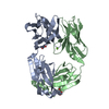
- PDB-8db4: Crystal structure of the peanut allergen Ara h 2 bound by two neu... -
+ Open data

- Basic information
| Entry | Database: PDB / ID: 8db4 | ||||||
|---|---|---|---|---|---|---|---|
| Title | Crystal structure of the peanut allergen Ara h 2 bound by two neutralizing antibodies 22S1 and 13T1 | ||||||
 Components Components |
| ||||||
 Keywords Keywords | ALLERGEN/IMMUNE SYSTEM / Antibody / immunotherapy / immunoglobulin / ALLERGEN-IMMUNE SYSTEM complex | ||||||
| Function / homology |  Function and homology information Function and homology information | ||||||
| Biological species |  Homo sapiens (human) Homo sapiens (human) | ||||||
| Method |  X-RAY DIFFRACTION / SYNCHROTRON / MOLECULAR REPLACEMENT / Resolution: 2.3 Å X-RAY DIFFRACTION / SYNCHROTRON / MOLECULAR REPLACEMENT / Resolution: 2.3 Å | ||||||
 Authors Authors | Min, J. / Pedersen, L.C. | ||||||
| Funding support |  United States, 1items United States, 1items
| ||||||
 Citation Citation | Journal: J.Clin.Invest. / Year: 2023 Title: Immunotherapy-induced neutralizing antibodies disrupt allergen binding and sustain allergen tolerance in peanut allergy. Authors: LaHood, N.A. / Min, J. / Keswani, T. / Richardson, C.M. / Amoako, K. / Zhou, J. / Marini-Rapoport, O. / Bernard, H. / Hazebrouck, S. / Shreffler, W.G. / Love, J.C. / Pomes, A. / Pedersen, L. ...Authors: LaHood, N.A. / Min, J. / Keswani, T. / Richardson, C.M. / Amoako, K. / Zhou, J. / Marini-Rapoport, O. / Bernard, H. / Hazebrouck, S. / Shreffler, W.G. / Love, J.C. / Pomes, A. / Pedersen, L.C. / Mueller, G.A. / Patil, S.U. | ||||||
| History |
|
- Structure visualization
| Structure viewer | Molecule: MolmilJmol/JSmol |
|---|
- Downloads & links
-Download
| PDBx/mmCIF format | 8db4.cif.gz | 408.4 KB | Display | PDBx/mmCIF format |
|---|---|---|---|---|
| PDB format | pdb8db4.ent.gz | 297.4 KB | Display | PDB format |
| PDBx/mmJSON format | 8db4.json.gz | Tree view | PDBx/mmJSON format | |
| Others |  Other downloads Other downloads |
-Validation report
| Arichive directory | https://data.pdbj.org/pub/pdb/validation_reports/db/8db4ftp://data.pdbj.org/pub/pdb/validation_reports/db/8db4 | HTTPS FTP |
|---|
-Related structure data

-Links
 PDBj
PDBj


- Assembly
| Deposited unit | 
| ||||||||||||
|---|---|---|---|---|---|---|---|---|---|---|---|---|---|
| 1 | 
| ||||||||||||
| 2 | 
| ||||||||||||
| Unit cell |
|
-Components
-Protein , 1 types, 2 molecules EF
| #5: Protein | Mass: 15660.163 Da / Num. of mol.: 2 Source method: isolated from a genetically manipulated source Source: (gene. exp.)  |
|---|
-Antibody , 4 types, 8 molecules AGBHCIDJ
| #1: Antibody | Mass: 24478.322 Da / Num. of mol.: 2 Source method: isolated from a genetically manipulated source Source: (gene. exp.) Homo sapiens (human) / Production host:   Cricetulus griseus (Chinese hamster) Cricetulus griseus (Chinese hamster)#2: Antibody | Mass: 23665.354 Da / Num. of mol.: 2 Source method: isolated from a genetically manipulated source Source: (gene. exp.) Homo sapiens (human) / Production host: Cricetulus griseus (Chinese hamster)#3: Antibody | Mass: 24291.121 Da / Num. of mol.: 2 Source method: isolated from a genetically manipulated source Source: (gene. exp.) Homo sapiens (human) / Production host: Cricetulus griseus (Chinese hamster)#4: Antibody | Mass: 23401.975 Da / Num. of mol.: 2 Source method: isolated from a genetically manipulated source Source: (gene. exp.) Homo sapiens (human) / Production host: Cricetulus griseus (Chinese hamster) |
|---|
-Non-polymers , 3 types, 317 molecules 


| #6: Chemical | ChemComp-ZN /  Mass: 65.409 Da / Num. of mol.: 9 / Source method: obtained synthetically / Formula: Zn Mass: 65.409 Da / Num. of mol.: 9 / Source method: obtained synthetically / Formula: Zn#7: Chemical | ChemComp-EDO / |  Mass: 62.068 Da / Num. of mol.: 1 / Source method: obtained synthetically / Formula: C2H6O2 Mass: 62.068 Da / Num. of mol.: 1 / Source method: obtained synthetically / Formula: C2H6O2#8: Water | ChemComp-HOH / | Mass: 18.015 Da / Num. of mol.: 307 / Source method: isolated from a natural source / Formula: H2O |
|---|
-Details
| Has ligand of interest | N |
|---|---|
| Has protein modification | Y |
-Experimental details
-Experiment
| Experiment | Method: X-RAY DIFFRACTION / Number of used crystals: 1 |
|---|
- Sample preparation
| Crystal | Density Matthews: 2.79 Å3/Da / Density % sol: 55.98 % |
|---|---|
| Crystal grow | Temperature: 277 K / Method: vapor diffusion, sitting drop / Details: 0.05 M zinc acetate, 20% PEG3350 |
-Data collection
| Diffraction | Mean temperature: 100 K / Serial crystal experiment: N |
|---|---|
| Diffraction source | Source: SYNCHROTRON / Site: APS / Beamline: 22-ID / Wavelength: 1 Å |
| Detector | Type: DECTRIS EIGER X 16M / Detector: PIXEL / Date: Apr 13, 2021 |
| Radiation | Protocol: SINGLE WAVELENGTH / Monochromatic (M) / Laue (L): M / Scattering type: x-ray |
| Radiation wavelength | Wavelength: 1 Å / Relative weight: 1 |
| Reflection | Resolution: 2.3→50 Å / Num. obs: 96622 / % possible obs: 91.4 % / Redundancy: 3.8 % / Biso Wilson estimate: 42.32 Å2 / CC1/2: 0.994 / CC star: 0.999 / Rmerge(I) obs: 0.078 / Rpim(I) all: 0.046 / Rrim(I) all: 0.091 / Χ2: 1.154 / Net I/σ(I): 20.8 |
| Reflection shell | Resolution: 2.3→2.34 Å / Redundancy: 3.7 % / Rmerge(I) obs: 0.772 / Mean I/σ(I) obs: 1.6 / Num. unique obs: 4898 / CC1/2: 0.795 / CC star: 0.941 / Rpim(I) all: 0.463 / Rrim(I) all: 0.902 / Χ2: 0.492 / % possible all: 93.1 |
- Processing
| Software |
| |||||||||||||||||||||||||||||||||||||||||||||||||||||||||||||||||||||||||||||||||||||||||||||||||||||||||||||||||||||||||||||||||||||||||||||||||||
|---|---|---|---|---|---|---|---|---|---|---|---|---|---|---|---|---|---|---|---|---|---|---|---|---|---|---|---|---|---|---|---|---|---|---|---|---|---|---|---|---|---|---|---|---|---|---|---|---|---|---|---|---|---|---|---|---|---|---|---|---|---|---|---|---|---|---|---|---|---|---|---|---|---|---|---|---|---|---|---|---|---|---|---|---|---|---|---|---|---|---|---|---|---|---|---|---|---|---|---|---|---|---|---|---|---|---|---|---|---|---|---|---|---|---|---|---|---|---|---|---|---|---|---|---|---|---|---|---|---|---|---|---|---|---|---|---|---|---|---|---|---|---|---|---|---|---|---|---|
| Refinement | Method to determine structure: MOLECULAR REPLACEMENT Starting model: PDB entry 4LLY & 3OB4 Resolution: 2.3→40.75 Å / SU ML: 0.3383 / Cross valid method: FREE R-VALUE / σ(F): 1.98 / Phase error: 31.1763 Stereochemistry target values: GeoStd + Monomer Library + CDL v1.2
| |||||||||||||||||||||||||||||||||||||||||||||||||||||||||||||||||||||||||||||||||||||||||||||||||||||||||||||||||||||||||||||||||||||||||||||||||||
| Solvent computation | Shrinkage radii: 0.9 Å / VDW probe radii: 1.11 Å / Solvent model: FLAT BULK SOLVENT MODEL | |||||||||||||||||||||||||||||||||||||||||||||||||||||||||||||||||||||||||||||||||||||||||||||||||||||||||||||||||||||||||||||||||||||||||||||||||||
| Displacement parameters | Biso mean: 54.53 Å2 | |||||||||||||||||||||||||||||||||||||||||||||||||||||||||||||||||||||||||||||||||||||||||||||||||||||||||||||||||||||||||||||||||||||||||||||||||||
| Refinement step | Cycle: LAST / Resolution: 2.3→40.75 Å
| |||||||||||||||||||||||||||||||||||||||||||||||||||||||||||||||||||||||||||||||||||||||||||||||||||||||||||||||||||||||||||||||||||||||||||||||||||
| Refine LS restraints |
| |||||||||||||||||||||||||||||||||||||||||||||||||||||||||||||||||||||||||||||||||||||||||||||||||||||||||||||||||||||||||||||||||||||||||||||||||||
| LS refinement shell |
|
