Movie
Movie Controller

[English] 日本語
 Yorodumi
- PDB-8d4c: beta-Arf1 mediated dimeric assembly of AP-1, Arf1, Nef complex wi... -
+ Open data

- Basic information
| Entry | Database: PDB / ID: 8d4c | ||||||||||||
|---|---|---|---|---|---|---|---|---|---|---|---|---|---|
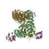
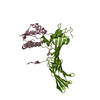
| Title | beta-Arf1 mediated dimeric assembly of AP-1, Arf1, Nef complex within lattice on MHC-I lipopeptide incorporated narrow membrane tubes | ||||||||||||
 Components Components |
| ||||||||||||
 Keywords Keywords | PROTEIN TRANSPORT / nef / AP / trafficking | ||||||||||||
| Function / homology |  Function and homology information Function and homology informationbasolateral protein secretion / synaptic vesicle budding from endosome / Lysosome Vesicle Biogenesis / positive regulation of natural killer cell degranulation / endosome to melanosome transport / protein trimerization / AP-1 adaptor complex / mitotic cleavage furrow ingression / trans-Golgi Network Vesicle Budding / negative regulation of glycoprotein biosynthetic process ...basolateral protein secretion / synaptic vesicle budding from endosome / Lysosome Vesicle Biogenesis / positive regulation of natural killer cell degranulation / endosome to melanosome transport / protein trimerization / AP-1 adaptor complex / mitotic cleavage furrow ingression / trans-Golgi Network Vesicle Budding / negative regulation of glycoprotein biosynthetic process / symbiont-mediated suppression of host antigen processing and presentation of peptide antigen via MHC class I / platelet dense granule organization / melanosome assembly / Glycosphingolipid transport / symbiont-mediated suppression of host antigen processing and presentation of peptide antigen via MHC class II / Golgi to lysosome transport / regulation of receptor internalization / Intra-Golgi traffic / Golgi to vacuole transport / Golgi Associated Vesicle Biogenesis / regulation of Arp2/3 complex-mediated actin nucleation / symbiont-mediated suppression of host autophagy / symbiont-mediated suppression of host apoptosis / Synthesis of PIPs at the Golgi membrane / clathrin-cargo adaptor activity / melanosome organization / MHC class II antigen presentation / GTP-dependent protein binding / thioesterase binding / Nef Mediated CD4 Down-regulation / positive regulation of natural killer cell mediated cytotoxicity / CD4 receptor binding / determination of left/right symmetry / dendritic spine organization / clathrin-coated vesicle / positive regulation of memory T cell activation / T cell mediated cytotoxicity directed against tumor cell target / long-term synaptic depression / positive regulation of CD8-positive, alpha-beta T cell activation / CD8-positive, alpha-beta T cell activation / positive regulation of CD8-positive, alpha-beta T cell proliferation / COPI-dependent Golgi-to-ER retrograde traffic / antigen processing and presentation of endogenous peptide antigen via MHC class I via ER pathway, TAP-dependent / TAP complex binding / Lysosome Vesicle Biogenesis / clathrin binding / antigen processing and presentation of exogenous peptide antigen via MHC class I / Golgi medial cisterna / Golgi Associated Vesicle Biogenesis / CD8 receptor binding / Synthesis of PIPs at the plasma membrane / cell leading edge / protection from natural killer cell mediated cytotoxicity / host cell Golgi membrane / beta-2-microglobulin binding / endoplasmic reticulum exit site / TAP binding / MHC class I protein binding / intracellular copper ion homeostasis / detection of bacterium / antigen processing and presentation of endogenous peptide antigen via MHC class Ib / antigen processing and presentation of endogenous peptide antigen via MHC class I via ER pathway, TAP-independent / kinesin binding / synaptic vesicle endocytosis / protein targeting / COPI-mediated anterograde transport / collagen binding / T cell receptor binding / viral life cycle / vesicle-mediated transport / Neutrophil degranulation / regulation of calcium-mediated signaling / clathrin-coated pit / MHC class II antigen presentation / Gene and protein expression by JAK-STAT signaling after Interleukin-12 stimulation / Nef mediated downregulation of MHC class I complex cell surface expression / cytoplasmic vesicle membrane / trans-Golgi network membrane / Endosomal/Vacuolar pathway / T cell mediated cytotoxicity / small monomeric GTPase / sarcomere / Antigen Presentation: Folding, assembly and peptide loading of class I MHC / lumenal side of endoplasmic reticulum membrane / kidney development / trans-Golgi network / peptide antigen assembly with MHC class I protein complex / ER to Golgi transport vesicle membrane / MHC class I peptide loading complex / positive regulation of T cell cytokine production / antigen processing and presentation of endogenous peptide antigen via MHC class I / MHC class I protein complex / intracellular protein transport / cellular response to virus / SH3 domain binding / positive regulation of T cell mediated cytotoxicity / recycling endosome / virion component / peptide antigen binding / positive regulation of type II interferon production Similarity search - Function | ||||||||||||
| Biological species |  Homo sapiens (human) Homo sapiens (human)  Human immunodeficiency virus 1 Human immunodeficiency virus 1 | ||||||||||||
| Method | ELECTRON MICROSCOPY / subtomogram averaging / cryo EM / Resolution: 9.3 Å | ||||||||||||
 Authors Authors | Hooy, R.M. / Hurley, J.H. | ||||||||||||
| Funding support |  United States, 3items United States, 3items
| ||||||||||||
 Citation Citation | Journal: Sci Adv / Year: 2022 Title: Self-assembly and structure of a clathrin-independent AP-1:Arf1 tubular membrane coat. Authors: Richard M Hooy / Yuichiro Iwamoto / Dan A Tudorica / Xuefeng Ren / James H Hurley / Abstract: The adaptor protein (AP) complexes not only form the inner layer of clathrin coats but also have clathrin-independent roles in membrane traffic whose mechanisms are unknown. HIV-1 Nef hijacks AP-1 to ...The adaptor protein (AP) complexes not only form the inner layer of clathrin coats but also have clathrin-independent roles in membrane traffic whose mechanisms are unknown. HIV-1 Nef hijacks AP-1 to sequester major histocompatibility complex class I (MHC-I), evading immune detection. We found that AP-1:Arf1:Nef:MHC-I forms a coat on tubulated membranes without clathrin and determined its structure. The coat assembles via Arf1 dimer interfaces. AP-1-positive tubules are enriched in cells upon clathrin knockdown. Nef localizes preferentially to AP-1 tubules in cells, explaining how Nef sequesters MHC-I. Coat contact residues are conserved across Arf isoforms and the Arf-dependent AP complexes AP-1, AP-3, and AP-4. Thus, AP complexes can self-assemble with Arf1 into tubular coats without clathrin or other scaffolding factors. The AP-1:Arf1 coat defines the structural basis of a broader class of tubulovesicular membrane coats as an intermediate in clathrin vesicle formation from internal membranes and as an MHC-I sequestration mechanism in HIV-1 infection. | ||||||||||||
| History |
|
- Structure visualization
| Structure viewer | Molecule: MolmilJmol/JSmol |
|---|
- Downloads & links
-Download
| PDBx/mmCIF format | 8d4c.cif.gz | 678.1 KB | Display | PDBx/mmCIF format |
|---|---|---|---|---|
| PDB format | pdb8d4c.ent.gz | Display | PDB format | |
| PDBx/mmJSON format | 8d4c.json.gz | Tree view | PDBx/mmJSON format | |
| Others |  Other downloads Other downloads |
-Validation report
| Arichive directory | https://data.pdbj.org/pub/pdb/validation_reports/d4/8d4cftp://data.pdbj.org/pub/pdb/validation_reports/d4/8d4c | HTTPS FTP |
|---|
-Related structure data
| Related structure data | 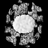 27181MC 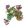 7ux3C  8d4dC  8d4eC  8d4fC  8d4gC  8d9rC  8d9sC  8d9tC  8d9uC  8d9vC  8d9wC C: citing same article ( M: map data used to model this data |
|---|---|
| Similar structure data |


-Links
 PDBj
PDBj


- Assembly
| Deposited unit | 
|
|---|---|
| 1 |
|
-Components
-Protein , 2 types, 8 molecules CHDFNLKI
| #1: Protein | Mass: 20590.547 Da / Num. of mol.: 4 Source method: isolated from a genetically manipulated source Details: N-terminal myristoylation / Source: (gene. exp.) Homo sapiens (human) / Gene: ARF1 / Production host:  #2: Protein | Mass: 24154.049 Da / Num. of mol.: 4 Source method: isolated from a genetically manipulated source Details: N-terminal myristoylation / Source: (gene. exp.) Human immunodeficiency virus 1 / Gene: nef / Production host: |
|---|
-Protein/peptide , 1 types, 2 molecules YP
| #3: Protein/peptide | Mass: 4139.429 Da / Num. of mol.: 2 / Mutation: T345S, S349G, G355S, C363A Source method: isolated from a genetically manipulated source Details: Conjugated to lipid maleimide via N-terminal cysteine Source: (gene. exp.) Homo sapiens (human) / Gene: HLA-A, HLAA / Production host: |
|---|
-AP-1 complex subunit ... , 4 types, 8 molecules BAGEMJSO
| #4: Protein | Mass: 104605.266 Da / Num. of mol.: 2 / Mutation: K359R, E476K Source method: isolated from a genetically manipulated source Source: (gene. exp.) Homo sapiens (human) / Gene: AP1B1, ADTB1, BAM22, CLAPB2 / Production host: #5: Protein | Mass: 68062.891 Da / Num. of mol.: 2 Source method: isolated from a genetically manipulated source Source: (gene. exp.) #6: Protein | Mass: 48606.730 Da / Num. of mol.: 2 Source method: isolated from a genetically manipulated source Source: (gene. exp.) #7: Protein | Mass: 18305.273 Da / Num. of mol.: 2 Source method: isolated from a genetically manipulated source Source: (gene. exp.) Homo sapiens (human) / Gene: AP1S3 / Production host: |
|---|
-Non-polymers , 2 types, 8 molecules 

| #8: Chemical | ChemComp-GTP /  Mass: 523.180 Da / Num. of mol.: 4 / Source method: obtained synthetically / Formula: C10H16N5O14P3 / Feature type: SUBJECT OF INVESTIGATION / Comment: GTP, energy-carrying molecule*YM Mass: 523.180 Da / Num. of mol.: 4 / Source method: obtained synthetically / Formula: C10H16N5O14P3 / Feature type: SUBJECT OF INVESTIGATION / Comment: GTP, energy-carrying molecule*YM#9: Chemical | ChemComp-MG /  Mass: 24.305 Da / Num. of mol.: 4 / Source method: obtained synthetically / Formula: Mg / Feature type: SUBJECT OF INVESTIGATION Mass: 24.305 Da / Num. of mol.: 4 / Source method: obtained synthetically / Formula: Mg / Feature type: SUBJECT OF INVESTIGATION |
|---|
-Details
| Has ligand of interest | Y |
|---|
-Experimental details
-Experiment
| Experiment | Method: ELECTRON MICROSCOPY |
|---|---|
| EM experiment | Aggregation state: PARTICLE / 3D reconstruction method: subtomogram averaging |
- Sample preparation
| Component |
| |||||||||||||||||||||||||
|---|---|---|---|---|---|---|---|---|---|---|---|---|---|---|---|---|---|---|---|---|---|---|---|---|---|---|
| Source (natural) |
| |||||||||||||||||||||||||
| Source (recombinant) |
| |||||||||||||||||||||||||
| Buffer solution | pH: 7.2 Details: HEPES/KOAc concentrated stocks are diluted to their final concentrations then pH'd to 7.2 with KOH prior to use in experiments. | |||||||||||||||||||||||||
| Buffer component |
| |||||||||||||||||||||||||
| Specimen | Conc.: 0.2 mg/ml / Embedding applied: NO / Shadowing applied: NO / Staining applied: NO / Vitrification applied: YES | |||||||||||||||||||||||||
| Specimen support | Grid type: EMS Lacey Carbon | |||||||||||||||||||||||||
| Vitrification | Instrument: FEI VITROBOT MARK IV / Cryogen name: ETHANE / Humidity: 100 % / Chamber temperature: 298 K Details: 60 second wait, 3-5 second blot, 597 filter paper, 0.5 second drain. Sample was supplemented with 10nm BSA-gold fiducials. 3.5ul of the mixture was double-side blotted. |
- Electron microscopy imaging
| Experimental equipment |  Model: Titan Krios / Image courtesy: FEI Company |
|---|---|
| Microscopy | Model: FEI TITAN KRIOS |
| Electron gun | Electron source:  FIELD EMISSION GUN / Accelerating voltage: 300 kV / Illumination mode: FLOOD BEAM FIELD EMISSION GUN / Accelerating voltage: 300 kV / Illumination mode: FLOOD BEAM |
| Electron lens | Mode: BRIGHT FIELD / Nominal magnification: 42000 X / Nominal defocus max: 4500 nm / Nominal defocus min: 1500 nm / Cs: 2.7 mm / Alignment procedure: COMA FREE |
| Specimen holder | Cryogen: NITROGEN / Specimen holder model: FEI TITAN KRIOS AUTOGRID HOLDER |
| Image recording | Average exposure time: 3 sec. / Electron dose: 3 e/Å2 / Avg electron dose per subtomogram: 123 e/Å2 / Film or detector model: GATAN K3 BIOQUANTUM (6k x 4k) / Num. of grids imaged: 1 Details: Tilt images were collected in movie-mode. Each movie/tilt consisted of 3-4 frames each |
| EM imaging optics | Energyfilter slit width: 25 eV |
| Image scans | Width: 5760 / Height: 4092 |
- Processing
| Software |
| ||||||||||||||||||||||||||||||||
|---|---|---|---|---|---|---|---|---|---|---|---|---|---|---|---|---|---|---|---|---|---|---|---|---|---|---|---|---|---|---|---|---|---|
| EM software |
| ||||||||||||||||||||||||||||||||
| Image processing | Details: The images were gain-normalized | ||||||||||||||||||||||||||||||||
| CTF correction | Details: CTF was estimated on a per-tilt basis in IMOD (4.11) using CTFPLOTTER. The results were used as input to NOVACTF during 3DCTF correction. Type: PHASE FLIPPING ONLY | ||||||||||||||||||||||||||||||||
| Symmetry | Point symmetry: C2 (2 fold cyclic) | ||||||||||||||||||||||||||||||||
| 3D reconstruction | Resolution: 9.3 Å / Resolution method: FSC 0.143 CUT-OFF / Num. of particles: 7004 / Symmetry type: POINT | ||||||||||||||||||||||||||||||||
| EM volume selection | Details: Tubes were annotated by tracing the center of the tube in Dynamo and recording the average apparent diameter. Initial subtomogram positions were picked using uniform radial and axial sampling. Num. of tomograms: 31 / Num. of volumes extracted: 61864 / Reference model: Reference-free | ||||||||||||||||||||||||||||||||
| Atomic model building | Protocol: RIGID BODY FIT | ||||||||||||||||||||||||||||||||
| Atomic model building | 3D fitting-ID: 1 / Source name: PDB / Type: experimental model
| ||||||||||||||||||||||||||||||||
| Refinement | Cross valid method: NONE Stereochemistry target values: GeoStd + Monomer Library + CDL v1.2 | ||||||||||||||||||||||||||||||||
| Displacement parameters | Biso mean: 32.79 Å2 | ||||||||||||||||||||||||||||||||
| Refine LS restraints |
|