 Movie
Movie Controller

[English] 日本語
 Yorodumi
- PDB-8cvb: Structure of Hyoscyamine 6-beta Hydroxylase in complex with iron,... -
+ Open data

- Basic information
| Entry | Database: PDB / ID: 8cvb | ||||||
|---|---|---|---|---|---|---|---|
| Title | Structure of Hyoscyamine 6-beta Hydroxylase in complex with iron, 2-oxoglutarate, and 6-OH-hyoscyamine | ||||||
 Components Components | Hyoscyamine 6-beta-hydroxylase | ||||||
 Keywords Keywords | OXIDOREDUCTASE / oxacyclase / scopolamine / epoxide | ||||||
| Function / homology |  Function and homology information Function and homology informationhyoscyamine (6S)-dioxygenase / hyoscyamine (6S)-dioxygenase activity / coumarin biosynthetic process / response to molecule of fungal origin / L-ascorbic acid binding / metal ion binding Similarity search - Function | ||||||
| Biological species |  Atropa belladonna (belladonna) Atropa belladonna (belladonna) | ||||||
| Method |  X-RAY DIFFRACTION / SYNCHROTRON / MOLECULAR REPLACEMENT / Resolution: 1.532 Å X-RAY DIFFRACTION / SYNCHROTRON / MOLECULAR REPLACEMENT / Resolution: 1.532 Å | ||||||
 Authors Authors | Wenger, E.S. / Boal, A.K. / Bollinger, J.M. / Krebs, C. | ||||||
| Funding support |  United States, 1items United States, 1items
| ||||||
 Citation Citation | Journal: J.Am.Chem.Soc. / Year: 2024 Title: Optimized Substrate Positioning Enables Switches in the C-H Cleavage Site and Reaction Outcome in the Hydroxylation-Epoxidation Sequence Catalyzed by Hyoscyamine 6 beta-Hydroxylase. Authors: Wenger, E.S. / Martinie, R.J. / Ushimaru, R. / Pollock, C.J. / Sil, D. / Li, A. / Hoang, N. / Palowitch, G.M. / Graham, B.P. / Schaperdoth, I. / Burke, E.J. / Maggiolo, A.O. / Chang, W.C. / ...Authors: Wenger, E.S. / Martinie, R.J. / Ushimaru, R. / Pollock, C.J. / Sil, D. / Li, A. / Hoang, N. / Palowitch, G.M. / Graham, B.P. / Schaperdoth, I. / Burke, E.J. / Maggiolo, A.O. / Chang, W.C. / Allen, B.D. / Krebs, C. / Silakov, A. / Boal, A.K. / Bollinger Jr., J.M. | ||||||
| History |
|
- Structure visualization
| Structure viewer | Molecule: MolmilJmol/JSmol |
|---|
- Downloads & links
-Download
| PDBx/mmCIF format | 8cvb.cif.gz | 95.1 KB | Display | PDBx/mmCIF format |
|---|---|---|---|---|
| PDB format | pdb8cvb.ent.gz | 65.7 KB | Display | PDB format |
| PDBx/mmJSON format | 8cvb.json.gz | Tree view | PDBx/mmJSON format | |
| Others |  Other downloads Other downloads |
-Validation report
| Arichive directory | https://data.pdbj.org/pub/pdb/validation_reports/cv/8cvbftp://data.pdbj.org/pub/pdb/validation_reports/cv/8cvb | HTTPS FTP |
|---|
-Related structure data
| Related structure data |  8cv8C  8cv9C 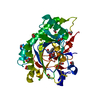 8cvaC 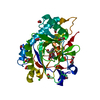 8cvcC  8cvdC 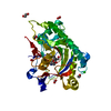 8cveC  8cvfC 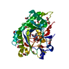 8cvgC  8cvhC  6ttmS S: Starting model for refinement C: citing same article ( |
|---|---|
| Similar structure data |
-Links
 PDBj
PDBj


- Assembly
| Deposited unit | 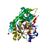
| ||||||||
|---|---|---|---|---|---|---|---|---|---|
| 1 |
| ||||||||
| Unit cell |
|
-Components
-Protein , 1 types, 1 molecules A
| #1: Protein | Mass: 42632.969 Da / Num. of mol.: 1 Source method: isolated from a genetically manipulated source Source: (gene. exp.) Atropa belladonna (belladonna) / Gene: AbH6H, h6h / Production host:  |
|---|
-Non-polymers , 7 types, 330 molecules 


| #2: Chemical | ChemComp-FE2 /  Mass: 55.845 Da / Num. of mol.: 1 / Source method: obtained synthetically / Formula: Fe / Feature type: SUBJECT OF INVESTIGATION Mass: 55.845 Da / Num. of mol.: 1 / Source method: obtained synthetically / Formula: Fe / Feature type: SUBJECT OF INVESTIGATION | ||||||||||
|---|---|---|---|---|---|---|---|---|---|---|---|
| #3: Chemical | ChemComp-EDO /  Mass: 62.068 Da / Num. of mol.: 5 / Source method: obtained synthetically / Formula: C2H6O2 Mass: 62.068 Da / Num. of mol.: 5 / Source method: obtained synthetically / Formula: C2H6O2#4: Chemical | ChemComp-FMT /  Mass: 46.025 Da / Num. of mol.: 9 / Source method: obtained synthetically / Formula: CH2O2 Mass: 46.025 Da / Num. of mol.: 9 / Source method: obtained synthetically / Formula: CH2O2#5: Chemical | ChemComp-AKG / |  Mass: 146.098 Da / Num. of mol.: 1 / Source method: obtained synthetically / Formula: C5H6O5 / Feature type: SUBJECT OF INVESTIGATION Mass: 146.098 Da / Num. of mol.: 1 / Source method: obtained synthetically / Formula: C5H6O5 / Feature type: SUBJECT OF INVESTIGATION#6: Chemical | ChemComp-OVR / ( |  Mass: 305.369 Da / Num. of mol.: 1 / Source method: obtained synthetically / Formula: C17H23NO4 / Feature type: SUBJECT OF INVESTIGATION Mass: 305.369 Da / Num. of mol.: 1 / Source method: obtained synthetically / Formula: C17H23NO4 / Feature type: SUBJECT OF INVESTIGATION#7: Chemical |  Mass: 87.620 Da / Num. of mol.: 2 / Source method: obtained synthetically / Formula: Sr Mass: 87.620 Da / Num. of mol.: 2 / Source method: obtained synthetically / Formula: Sr#8: Water | ChemComp-HOH / | Mass: 18.015 Da / Num. of mol.: 311 / Source method: isolated from a natural source / Formula: H2O |
-Details
| Has ligand of interest | Y |
|---|---|
| Has protein modification | N |
-Experimental details
-Experiment
| Experiment | Method: X-RAY DIFFRACTION / Number of used crystals: 1 |
|---|
- Sample preparation
| Crystal | Density Matthews: 2.15 Å3/Da / Density % sol: 42.95 % / Description: Bar |
|---|---|
| Crystal grow | Temperature: 293 K / Method: vapor diffusion, hanging drop / Details: PEG 3350, sodium formate, strontium chloride |
-Data collection
| Diffraction | Mean temperature: 100 K / Serial crystal experiment: N |
|---|---|
| Diffraction source | Source: SYNCHROTRON / Site: APS / Beamline: 21-ID-F / Wavelength: 0.97872 Å |
| Detector | Type: RAYONIX MX-300 / Detector: CCD / Date: Oct 10, 2020 |
| Radiation | Monochromator: C(111) / Protocol: SINGLE WAVELENGTH / Monochromatic (M) / Laue (L): M / Scattering type: x-ray |
| Radiation wavelength | Wavelength: 0.97872 Å / Relative weight: 1 |
| Reflection | Resolution: 1.53→50 Å / Num. obs: 54702 / % possible obs: 99.9 % / Redundancy: 7.6 % / CC1/2: 0.93 / Rmerge(I) obs: 0.046 / Rpim(I) all: 0.018 / Net I/σ(I): 34.8 |
| Reflection shell | Resolution: 1.53→1.56 Å / Redundancy: 6.5 % / Rmerge(I) obs: 0.371 / Mean I/σ(I) obs: 5 / Num. unique obs: 2733 / CC1/2: 0.93 / Rpim(I) all: 0.157 / % possible all: 99 |
- Processing
| Software |
| ||||||||||||||||||||||||||||||||||||||||||||||||||||||||||||||||||||||||||||||||||||||||||||||||||||||||||||||||||||||||||||||||||||||||||||||||||||||||||||||||
|---|---|---|---|---|---|---|---|---|---|---|---|---|---|---|---|---|---|---|---|---|---|---|---|---|---|---|---|---|---|---|---|---|---|---|---|---|---|---|---|---|---|---|---|---|---|---|---|---|---|---|---|---|---|---|---|---|---|---|---|---|---|---|---|---|---|---|---|---|---|---|---|---|---|---|---|---|---|---|---|---|---|---|---|---|---|---|---|---|---|---|---|---|---|---|---|---|---|---|---|---|---|---|---|---|---|---|---|---|---|---|---|---|---|---|---|---|---|---|---|---|---|---|---|---|---|---|---|---|---|---|---|---|---|---|---|---|---|---|---|---|---|---|---|---|---|---|---|---|---|---|---|---|---|---|---|---|---|---|---|---|---|
| Refinement | Method to determine structure: MOLECULAR REPLACEMENT Starting model: 6TTM Resolution: 1.532→43.61 Å / Cor.coef. Fo:Fc: 0.957 / Cor.coef. Fo:Fc free: 0.944 / SU B: 1.222 / SU ML: 0.046 / Cross valid method: FREE R-VALUE / ESU R: 0.077 / ESU R Free: 0.076 Details: Hydrogens have been added in their riding positions
| ||||||||||||||||||||||||||||||||||||||||||||||||||||||||||||||||||||||||||||||||||||||||||||||||||||||||||||||||||||||||||||||||||||||||||||||||||||||||||||||||
| Solvent computation | Ion probe radii: 0.8 Å / Shrinkage radii: 0.8 Å / VDW probe radii: 1.2 Å / Solvent model: MASK BULK SOLVENT | ||||||||||||||||||||||||||||||||||||||||||||||||||||||||||||||||||||||||||||||||||||||||||||||||||||||||||||||||||||||||||||||||||||||||||||||||||||||||||||||||
| Displacement parameters | Biso mean: 14.553 Å2
| ||||||||||||||||||||||||||||||||||||||||||||||||||||||||||||||||||||||||||||||||||||||||||||||||||||||||||||||||||||||||||||||||||||||||||||||||||||||||||||||||
| Refinement step | Cycle: LAST / Resolution: 1.532→43.61 Å
| ||||||||||||||||||||||||||||||||||||||||||||||||||||||||||||||||||||||||||||||||||||||||||||||||||||||||||||||||||||||||||||||||||||||||||||||||||||||||||||||||
| Refine LS restraints |
| ||||||||||||||||||||||||||||||||||||||||||||||||||||||||||||||||||||||||||||||||||||||||||||||||||||||||||||||||||||||||||||||||||||||||||||||||||||||||||||||||
| LS refinement shell |
|
